Figure 2.
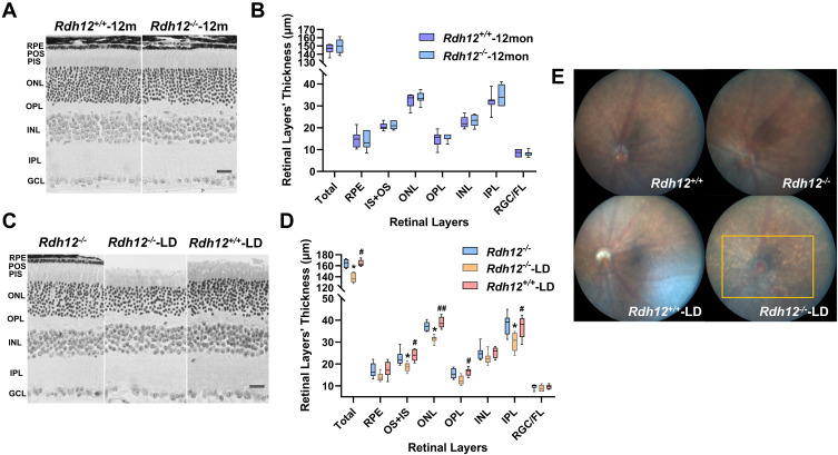
Photoreceptor degeneration was induced by bright light in Rdh12−/− mice. (A) Montage of cross-sections of the retina from Rdh12−/− and Rdh12+/+ mice that raised in 12h dark/12h light conditions for 1 year. (B) Quantify the thickness of different layers in Rdh12−/− and Rdh12+/+ mice retina, which were measured at 1000 µm from the optic nerve head. (C) Montage of cross-sections of the retina from Rdh12−/−, Rdh12−/−-LD and Rdh12+/+-LD mice, which exposed to light for 48 h and then dark-adapted for 24h. The ages of mice were 6–8 weeks. (D) Quantify the thickness of different layers in Rdh12−/−, Rdh12−/−-LD and Rdh12+/+-LD mice retinas, which were measured at 1000 µm from the optic nerve head. (E) Fundus camera detected fundus appearance. Results were presented as median (min - max). Scale bar, 20 µm. n=8–12 per group. *P < 0.05 vs Rdh12−/− group. #P < 0.05; ##P < 0.01 vs Rdh12−/−-LD group.
Abbreviations: RGC/FL, retinal ganglion cell/fiber layer; IPL, inner plexiform layer; INL, inner nuclear layer; OPL, outer plexiform layer; ONL, outer nuclear layer; IS, inner segment; OS, outer segment; RPE, retinal pigment epithelium.

