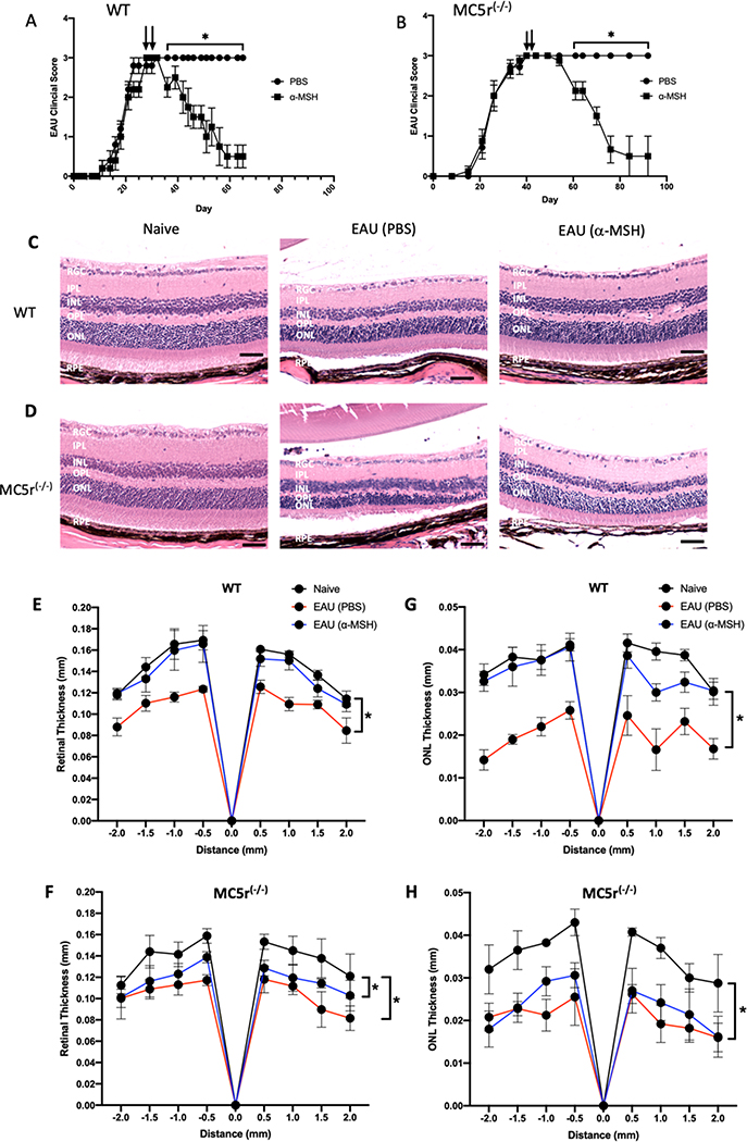
Figure 1. The effects of α-MSH-treatment on EAU in MC5r(−/−) mice.
EAU was induced in Wildtype and MC5r(−/−) mice and treated with α-MSH when clinical scores reached the chronic phase of inflammation (clinical score = 3) on Day 30 for WT mice and Day 40 for the MC5r(−/−) with a second injection of α-MSH 2 days later. The mean EAU scores ± SEM of each treated group (N = 10) of A) WT and B) MC5r(−/−) mice are presented over time. Representative retinal sections on Day 65 of WT mice and Day 90 of MC5r(−/−) mice were stained and presented. C) The retinas of α-MSH treated EAU mice (EAU (α-MSH)) appeared the same as retinas of naive mouse eyes. The vehicle injected (PBS) EAU retina was the thinnest with cell loss in both inter-nuclear layer and outer-nuclear layer. D) Both α-MSH and PBS injected MC5r(−/−) EAU mice had retinas that were thinner than the naive mouse retina with about half the photoreceptor outer-segment layer lost in the PBS-injected MC5r(−/−) EAU mouse retinas. The size bar equals 50μm. RGC – Retinal Ganglion Cells Layer; IPL – Inner Plexiform Layer; INL – Inner Nuclear Layer; OPL – Outer Plexiform Layer; ONL – Outer Nuclear Layer; RPE – Retinal Pigment Epithelium. The thickness of retina (E, F) and ONL (G, H) (N=5 each) were measured by ImageJ and presented as mean (mm) of thickness ± SEM at 0.5 mm intervals from the optic nerve head. There is no statistical difference between WT and α-MSH-treated EAU-WT retinal and ONL thickness (E, G); whereas there were (* P ≤ 0.001) significant loss of retinal and ONL thickness in the untereated and α-MSH-treated EAU MC5r(−/−) mice.

