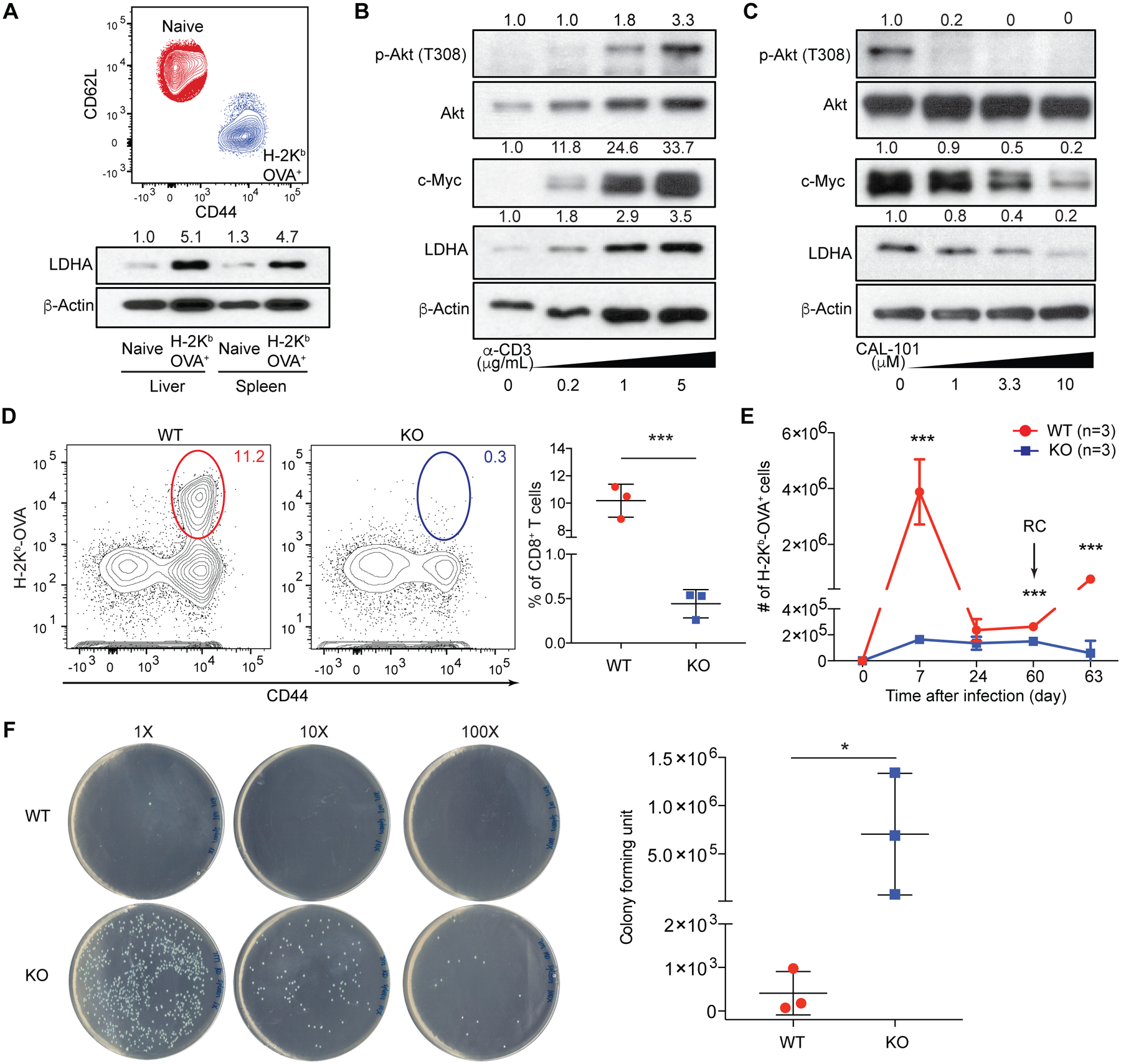
Fig. 1. PI3K-dependent expression of LDHA in CD8+ effector T cells is essential for antibacterial immunity.

(A) Flow cytometric analysis of CD44 and CD62L expression in splenic naïve and H-2Kb-OVA+ CD8+ T cells 7 days post LM-OVA infection. Immunoblotting and normalized expression of LDHA to β-actin. (B) Naïve CD8+ T cells were stimulated with anti-CD3 in the presence of anti-CD28, and IL-2. Immunoblotting and normalized expression of p-Akt (T308) to Akt as well as c-Myc and LDHA to β-actin. (C) Naïve CD8+ T cells were stimulated with anti-CD3, anti-CD28, and IL-2 in the presence of the PI3K inhibitor, CAL-101. Immunoblotting and normalized expression of p-Akt (T308) to Akt as well as c-Myc and LDHA to β-actin. (D to F) Ldhafl/fl (wild-type, WT) and Tbx21CreLdhafl/fl (knockout, KO) mice were infected followed or not by re-challenge (RC) with LM-OVA. (D) Representative flow cytometry plots and frequencies of splenic H-2Kb-OVA+ CD8+ T cells 7 days post LM-OVA infection. (E) H-2Kb-OVA+ CD8+ T cells were enumerated 7, 24, 60 days post-infection, and day-3 post-secondary infection. (F) Splenic bacterial burden from day-3-RC mice. Data are representative of three (A to C) and two (D to F, n= 3 per genotype, mean ± SD) independent experiments. Unpaired t tests for the measurements between the two groups (D and F) and multiple t tests between the two groups (E): *p<0.05 and ***p<0.001.
