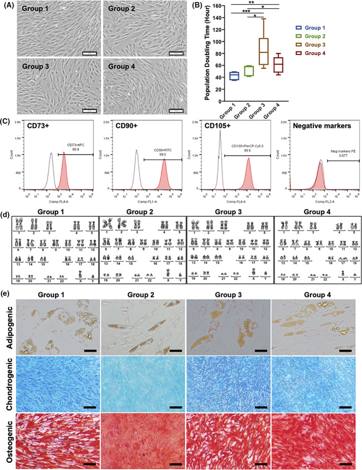
FIGURE 2.

Characterization of autologous bone marrow‐derived mesenchymal stem cells (BM‐MSCs) isolated from the control group (group 1) and from patients with type 2 diabetes mellitus (T2DM) (duration of T2DM: group 2: <5 years, group 3:5‐10 years, and group 4: >10 years). A, Typical morphology of BM‐MSCs cultured under xeno‐ and serum‐free culture conditions. These cells were plastic‐adherent, spindle‐shaped, and fibroblast‐like. B, The population doubling time (hours) of BM‐MSCs indicated a significant difference in the growth rates of groups 3 and 4 compared with those of group 1 (control group) and group 2. C, The flow cytometry analysis of mesenchymal stem cell surface markers, including the positive markers CD73, CD90, and CD105, and the absence of hematopoietic markers (CD34, CD45, CD14 or CD11b, CD79α or CD19, and HLA‐DR) confirmed that all BM‐MSCs expressed more than 95% of positive markers and less than 2% of negative markers. D, A normal karyotype was maintained in all cells cultured under xeno‐ and serum‐free conditions. E, All BM‐MSCs from the four groups were able to differentiate into adipocytes, chondrocytes, and osteoblasts. Scale bar: 100 μm. APC, allophycocyanin; Cy5.5, cyanine dyes 5.5; FITC, fluorescein isothiocyanate; HLA‐DR, human leukocyte antigen ‐ DR isotype; PE, phycoerythrin; PerCP, peridinin chlorophyll protein complex; *P<.05; **P<0.01; ***P<0.001
