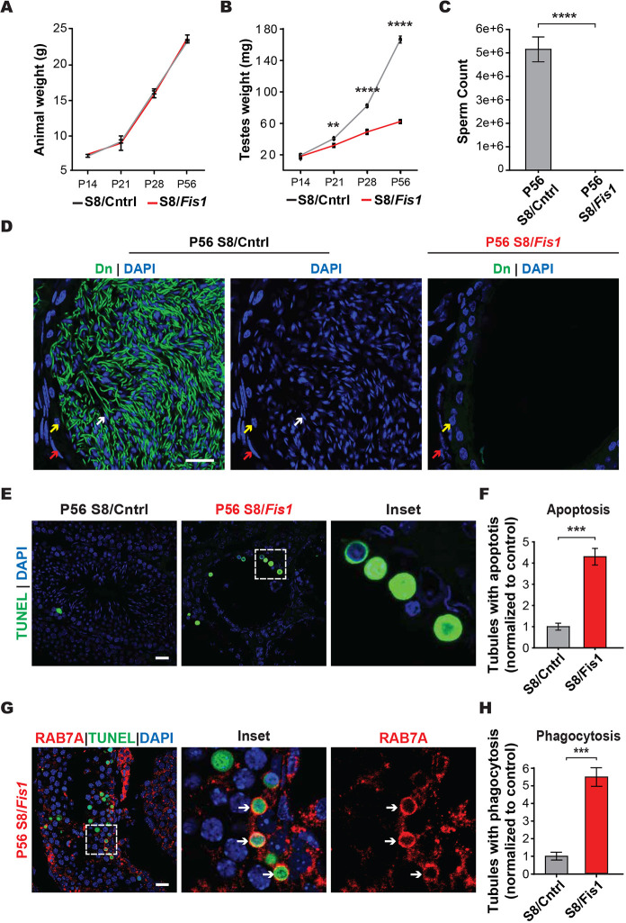
Fig. 1.
Fis1 is required for spermatogenesis. (A) Longitudinal analysis of animal weight. (B) Longitudinal analysis of testis weight. (C) Epididymal sperm count. Spermatozoa from both caudal epididymides were counted. (D) Histological analysis of caudal epididymis sections. Nuclei were stained with DAPI (blue), and mitochondria were labeled with mito-Dendra2 (Dn) (green). Note that no spermatozoa were present in the mutant sample. Red arrows, smooth muscle cells; yellow arrows, basal cells; white arrows; spermatozoa. Scale bar: 20 µm. (E) TUNEL staining (green) to detect apoptotic cells in testis sections. Scale bar: 20 µm. (F) Quantification of the number of tubules containing one or more apoptotic germ cells, normalized to control. (G) Histological analysis of apoptotic cells. Apoptotic cells (green) are surrounded by a RAB7A-positive structure (red), which is likely a Sertoli cell phagosome (white arrows). Scale bar: 20 µm. (H) Quantification of the number of tubules containing one or more RAB7A phagosomes, normalized to control. All data are from adult (P56) mice. Data are represented as mean±s.e.m. ****P≤0.0001; ***P≤0.001. For statistical tests used, see the Materials and Methods section.

