Fig. 2.
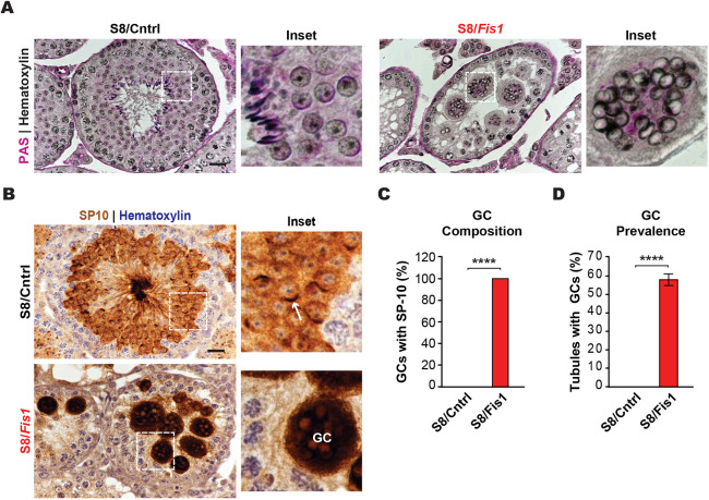
Germ cell Fis1 deletion results in multinucleated spermatid giant cells. (A) PAS staining of adult testis sections, counterstained with Hematoxylin. Note the large, multinucleated GCs in S8/Fis1 testes. Scale bar: 20 µm. (B) Immunohistochemical staining of testis sections with an antibody against the spermatid-specific SP-10 protein. The acrosome in a control round spermatid is indicated by a white arrow. Spermatids in mutant GCs lack acrosomes and stain intensely for SP-10. Scale bar: 20 µm. (C) Quantification of the number of GCs with SP-10 reactivity. (D) Quantification showing the prevalence of spermatid GCs in Fis1 mutants. Control testes never exhibit GCs. All data are from adult (P56) mice. Data are represented as mean±s.e.m. ****P≤0.0001. For statistical tests used, see the Materials and Methods section.

