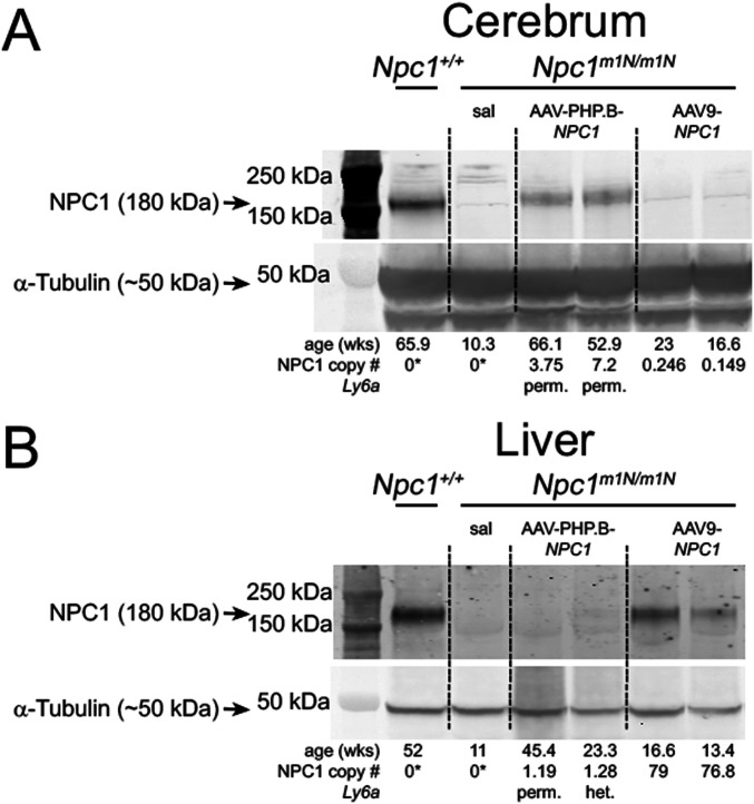
Figure 4. NPC1 protein levels correspond with differential transduction efficiency of adeno-associated virus (AAV)-PHP.B-NPC1 and AAV9-NPC1 vectors.
(A, B) Western blots were used to confirm the presence or absence of NPC1 protein in cerebrum and liver tissue. The Npc1m1N/m1N model is a null, thus the only NPC1 protein present would arise from the transduced vectors. Age in weeks and NPC1 copy number for each mouse is under α-tubulin loading control. (A) In cerebrum, NPC1 protein was detectable only in the longest surviving AAV-PHP.B-NPC1–treated Npc1m1N/m1N and Npc1+/+ mice, and was not detected in AAV9-NPC1–treated Npc1m1N/m1N mice. (B) Conversely, analysis of liver showed that NPC1 protein was present in most AAV9-NPC1–treated Npc1m1N/m1N mice, but only rarely in Npc1m1N/m1N mice receiving AAV-PHP.B-NPC1. *Droplet digital PCR assayed only human NPC1, not murine NPC1, hence zero values for non-gene therapy treated mice. Full unedited gels for Fig 4: Please see green outlines below for cerebrum and blue outlines below for liver to denote the portion of the gel used in Fig 4. Each gel was co-labeled with NPC1 and α-tubulin and different secondaries were used for each primary antibody.
Source data are available for this figure.

