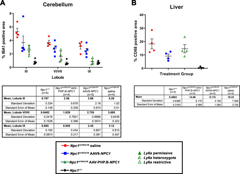
Figure S7. Quantification of percent positive area of macrophages versus total area assessed in cerebellum and liver reveals no significant differences in pathology, other than the expected increase in Npc1m1N/m1N saline-injected mice versus Npc1+/+ mice.
(A) Quantification of anti-IBA1 staining (microglial marker) in cerebellar lobules III, VI/VII, and IX suggests modestly reduced pathology in the Npc1m1N/m1N mice treated with either adeno-associated virus (AAV)9-NPC1 or AAV-PHP.B-NPC1 compared with saline-injected Npc1m1N/m1N mice, with AAV-PHP.B-NPC1 trending towards greater pathology reduction. (B) Quantification of anti-CD68 staining (macrophage marker) suggests only modest reduction in liver pathology of Npc1m1N/m1N mice after treatment with either AAV9-NPC1 or AAV-PHP.B-NPC1 compared with saline-injected Npc1m1N/m1N mice, with a trend towards greater pathology reduction by AAV9-NPC1.

