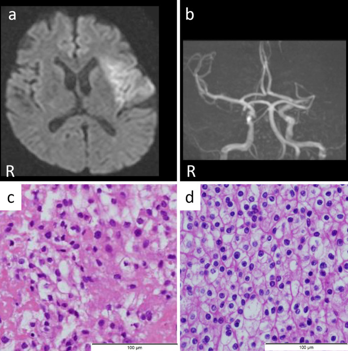
Figure.
(a) Axial diffusion-weighted image (DWI, 1.5 T; B value 1,000 s/mm2, TR 6,000 ms, TE 100 ms) shows a hyperintense lesion in the left insular cortex and middle cerebral artery cortex. (b) MRA shows left middle cerebral artery occlusion. (c) Retrieved thrombus (×200). Hematoxylin and Eosin staining shows a solid mass growing in the fibrin-based thrombus. (d) Microscopic image of the renal cell carcinoma (×200). The pathological findings are closely consistent with the retrieved thrombus.

