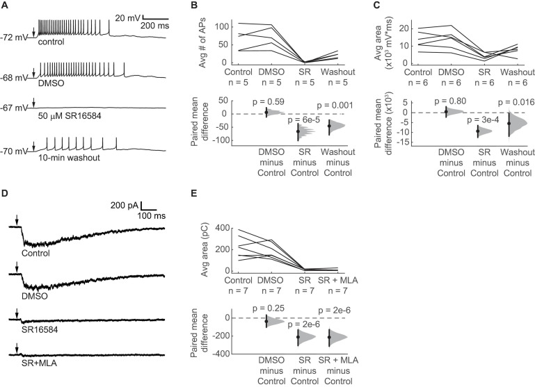
FIGURE 6.
ACh-induced depolarization of VIP neurons is predominately mediated by α3β4* nAChRs. (A) Example traces show that 50 μM SR16584, an α3β4* nAChR antagonist, blocked action potential firing and most of the depolarization elicited by 1 mM ACh puffs. Conditions from top to bottom: control, vehicle (1:1000 DMSO:ACSF), 50 μM SR16584, and 10-min washout (control ACSF). Arrows indicate the time of the ACh puffs, and voltages indicate resting membrane potential. (B) The average number of action potentials elicited by ACh was reduced to 0.64 ± 1.22 (mean ± SD) after bath application of SR16584 (LMM: treatment effect, F3,12 = 20.33, p = 5e-5, n = 5; control vs. DMSO, t12 = 0.55, p = 0.59; control vs. SR, t12 = –6.01, p = 6e-5; control vs. washout, t12 = –4.13, p = 0.001). (C) The average total depolarization was also significantly decreased by application of SR16584 (LMM: treatment effect, F3,15 = 10.90, p = 5e-4, n = 6; control vs. DMSO, t15 = 0.26, p = 0.80, control vs. SR, t15 = –4.64, p = 3e-4; control vs. washout, t15 = –2.71, p = 0.016). (D) Example traces show that 50 μM SR16584 blocked nearly all of the inward current elicited by 1 mM ACh puffs. Conditions from top to bottom: control, vehicle (1:1000 DMSO:ACSF), 50 μM SR16584, 50 μM SR16584 + 5 nM MLA. Arrows indicate the time of the ACh puffs. Holding potential was –60 mV. (E) Inward current elicited by ACh was decreased to 7.4 ± 6.2% (mean ± SD) of control by SR16584 (LMM: treatment effect, F3,18 = 27.5, p = 6e-7, n = 7; control vs. DMSO, t18 = –1.20, p = 0.25; control vs. SR, t18 = –6.93, p = 2e-6; control vs. SR + MLA, t18 = –7.01, p = 2e-6). (B,C,E) Horizontal dashed lines indicate the level of zero mean difference. Vertical lines on the paired mean difference points indicate 95% bootstrap confidence intervals.

