
Figure 1.
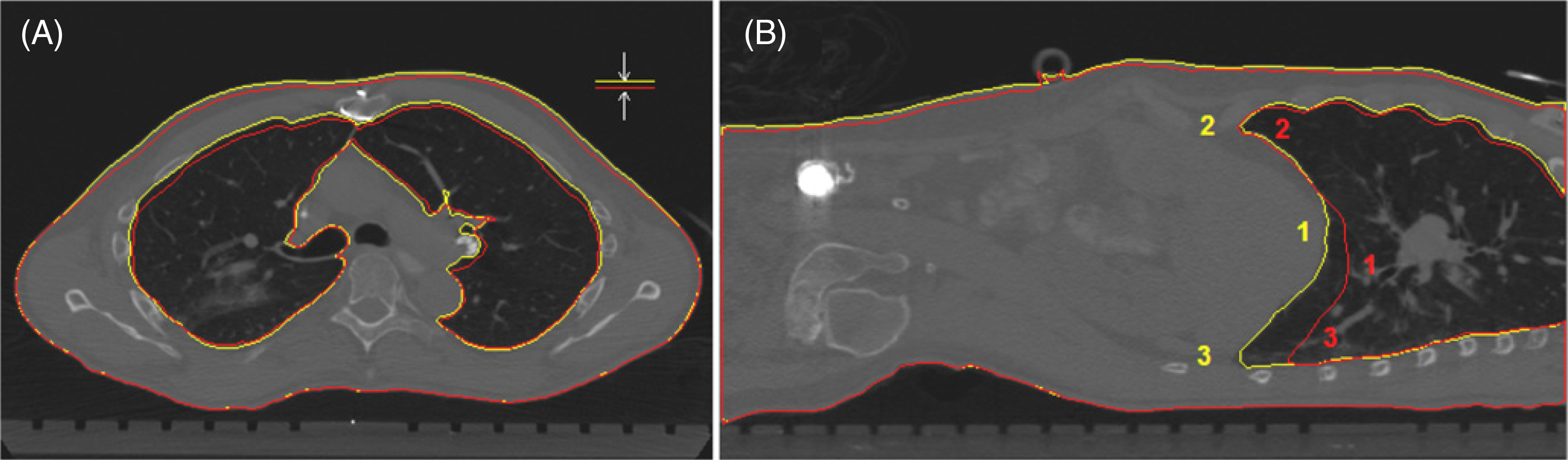
Demonstration of the similarity between thoracic surface height variation and anterior lung height variation. Torso and lung contours in full-exhalation (red) and full-inhalation (yellow) stage CTs of a patient in supine position. (A) The axial view also shows that lateral width variations of the lung and body are small. (B) The sagittal view illustrates the unevenness of diaphragm motion, as well as three locations (1, 2 and 3) that are used for calculating the position and displacement of the averaged diaphragm and individual points.