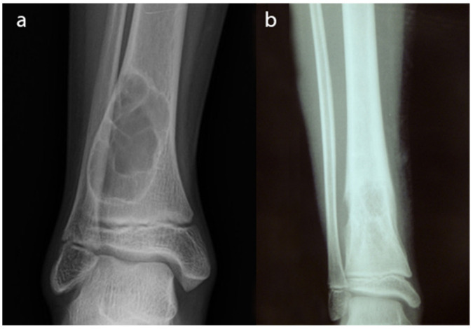
Fig. 1.

Radiographs of a non-ossifying fibroma in the distal tibia. This is a benign lesion. Well delineated borders and sclerotic edges can be appreciated. There is no cortical breakage and no soft-tissue mass; b) radiographs of an Ewing sarcoma of the distal tibia. This is a malignant bone lesion with undefined borders and a speckled pattern. Breakage of the cortical is well observed.
