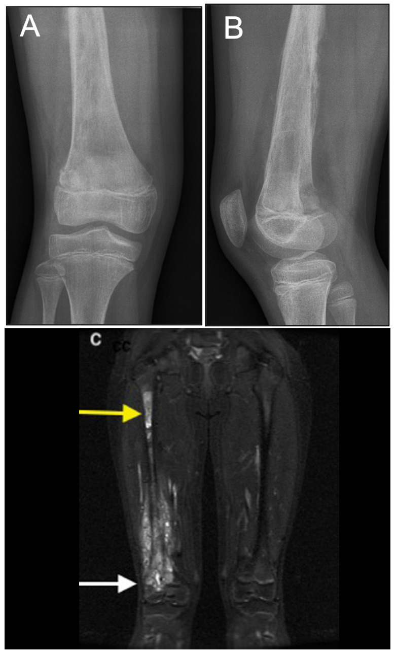
Fig. 4.

Right femur osteosarcoma in a 11-year-old girl: a) and b) on the radiographs the lesion cannot be well delimited; c) the MRI study reveals a large lesion with epiphyseal extension (white arrow) and a skip lesion in the proximal diaphysis (yellow arrow).
