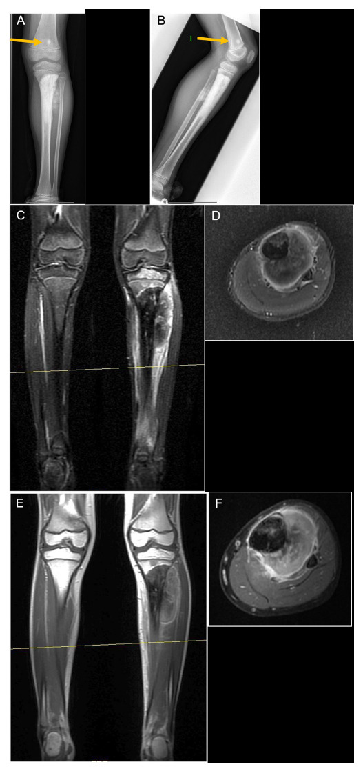
Fig. 5.

Left tibia osteosarcoma in a seven-year-old boy: a) and b) radiographs show an osteoblastic lesion in the proximal tibia, with a skip lesion in the distal femur (yellow arrow). Biopsy showed a high-grade osteosarcoma with osteoblastic predominance; c) and d) in short tau inversion recovery images of the lesion, it is difficult to distinguish between tumour and oedema; a) and f) T1 fat saturation images with contrast, where the tumour can be better delineated.
