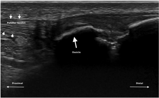
Figure 4.

Representative image of participant presenting with ossicle on ultrasound examination at follow-up. Participant reported minimal pain on palpation at clinical examination. Small arrows demonstrate the border of the patellar tendon, while the large arrow indicates the ossicle.
