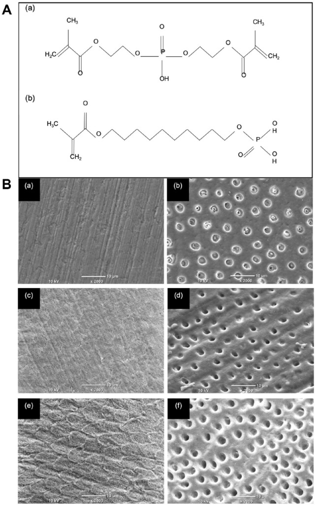
Figure 1.

The chemical structure of the phosphoric acid esters used in this study and the etch patterns produced on enamel and dentine. (A) The structure of (a) 10-methacryloyloxydecyl dihydrogenphosphate (10-MDP) and (b) bis[2-(methacryloyloxy) ethyl] phosphate (BMEP). (B) Scanning electron microscopy images of the self-etch primers on enamel (left column) and dentine (right column) using (a, b) CFSE, (c, d) BMEP15, and (e, f) BMEP40. A distinct etch pattern was obtained with BMEP40 on enamel (e), exposing the enamel prisms. A decrease in pH of the primers (right column, top to bottom) also increased the extent of demineralization, and the dentine tubules were enlarged (f).
