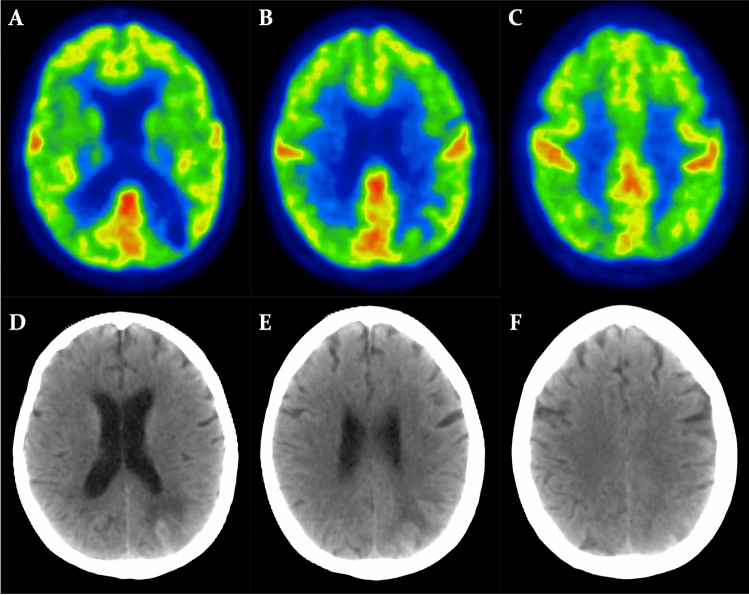
Fig. 4.
Brain FDG-PET findings in case 2. Matched axial brain FDG-PET (A,B,C) and CT scan (D,E,F) showing diffuse bilateral hypometabolism with frontal predominance. Additionally, a focal area of severe hypometabolism (A, B), resulting from a spontaneous parenchymal haematoma associated with subarachnoid haemorrhage (D, E), is observable in the left parietal lobe. FDG-PET [18F]FDG positron emission tomography. CT computed tomography

