Figure 1.

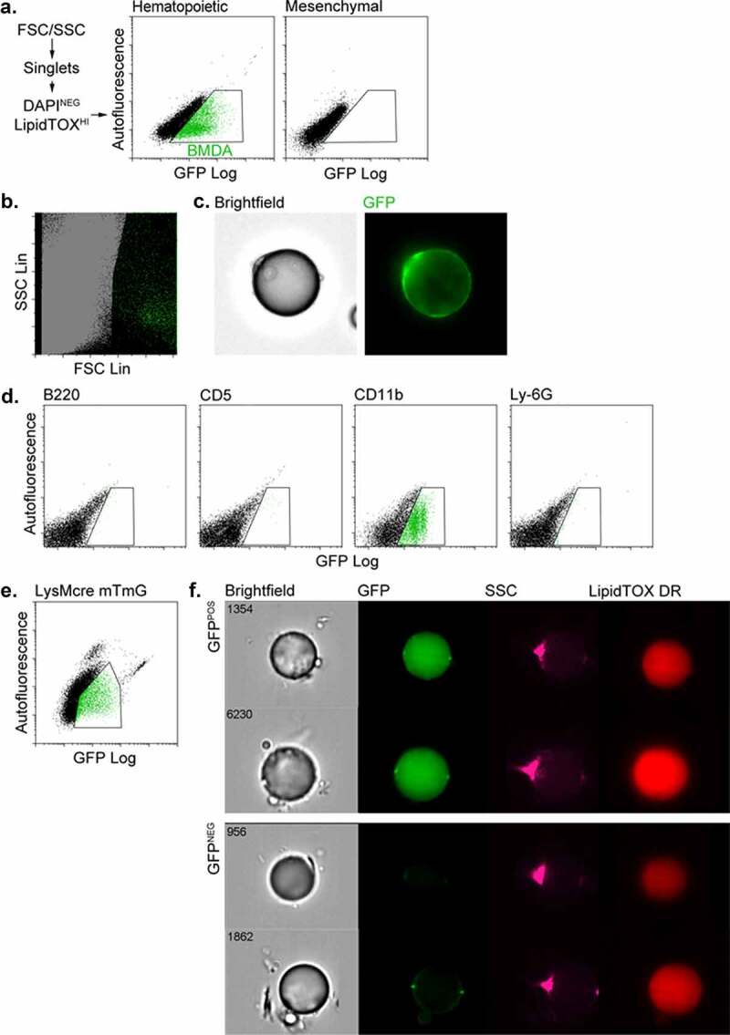
BMDAs are generated from HSCs via the myeloid lineage. (a) Competitive transplants were performed with either haematopoietic or mesenchymal progenitor cells constitutively expressing GFP, mixed with GFP-naïve carrier cells. After 8 weeks, free-floating adipocytes were prepared from adipose tissue from the transplanted mice and analysed by flow cytometry. GFPPOS BMDAs were detected in the adipocyte fraction of mice transplanted with GFP-labelled HSC (gated green events) but not labelled BM mesenchymal stem cells. (b) Backgating of the GFPPOS events in Figure 1a confirmed their large size (high forward scatter) and low internal complexity (low forward scatter) common to unilocular adipocytes. (c) Images of GFPPOS adipocytes from mice transplanted with GFPPOS HSC confirmed their identity as unilocular adipocytes with a single nucleus and cytosolic GFP fluorescence. (d) Haematopoietic sub-population cells were isolated from the BM of GFP-expressing mice with magnetic bead bearing antibodies to specific lineages (B220 = B lymphocyte, CD5 = T lymphocyte, CD11b = myeloid and Ly-6 G = granulocytes). The separated GFPPOS BM cells were combined with GFPNEG BM cells from wild type mice to ensure survival of recipients. After 8 weeks flow cytometry showed production of BMDAs only in mice transplanted with labelled (Cd11bPOS) myeloid cells. (e) Flow cytometry of free-floating adipocytes isolated from dual transgenic mice in which GFP expression (from an mTmG transgene) was controlled by the myeloid-specific lysozyme gene promoter (LysMcre). A population of GFPPOS adipocytes confirmed that some adipocytes were produced from the myeloid lineage. (f) Free-floating adipocytes were isolated from the LysMcre mTmG mice and subjected to imaging flow cytometry. Brightfield and fluorescence images confirmed the presence of GFPPOS unilocular (single LipidTOX stained lipid droplet) adipocytes in additional to GFPNEG adipocytes
