
Figure 8.
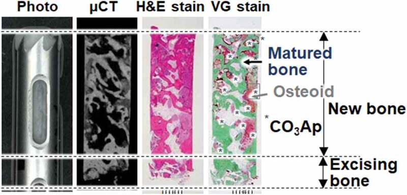
Photo, μ-CT scanning, H-E and VG staining of a histological specimen biopsied from a patient who underwent two-stage sinus floor augmentation for dental implantation [34]
Official websites use .gov
A
.gov website belongs to an official
government organization in the United States.
Secure .gov websites use HTTPS
A lock (
) or https:// means you've safely
connected to the .gov website. Share sensitive
information only on official, secure websites.
Photo, μ-CT scanning, H-E and VG staining of a histological specimen biopsied from a patient who underwent two-stage sinus floor augmentation for dental implantation [34]