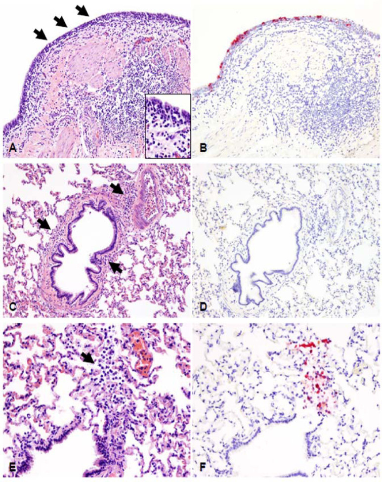
Figure 6. Histopathological lesions and SARS-CoV-2 antigen distribution in the lower respiratory tract of white-tailed deer at 4 days post-challenge (DPC).
The bronchial mucosa was characterized by segmental attenuation of the lining respiratory epithelium with loss of cilia, degeneration/necrosis of individual epithelial cells and neutrophil and lymphocyte transmigration, and a mixed lymphocytic and histiocytic infiltrate in the edematous lamina propria (A, arrows and inset). The epithelium lining of affected segments frequently contains viral antigen (B). In the remaining pulmonary parenchyma, bronchioles and blood vessels are delimited by perivascular and peribronchiolar lymphocytes, histiocytes and few neutrophils (C, arrows). Viral antigen is generally not detected (D). Rarely, sloughed and necrotic epithelial cells and few degenerate leukocytes lodged at the termini of respiratory bronchioles (E) contain intracytoplasmic viral antigen (F). H&E and Fast Red, 200X total magnification.

