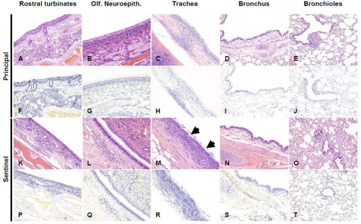
Figure 6. Histological lesions and SARS-CoV-2 antigen distribution in the upper and lower respiratory tract of principal infected and sentinel white-tailed deer at 18 DPC.
Rostral turbinates (A, F, K, P), olfactory neuroepithelium (B, G, L, Q), trachea (C, H, M, R), bronchus (D, I, N, S) and bronchioles (E, J, O, T). In principal infected deer, few lymphocytes were noted in the lamina propria of the rostral turbinates (A). The olfactory neuroepithelium was histologically unremarkable (B), and the tracheal and bronchial lamina propria were occasionally infiltrated by either dispersed or aggregates of mononuclear cells (C, D), which also encircled few bronchioles and pulmonary vessels (E). In sentinel deer, the lamina propria of rostral turbinates and sporadically subjacent to the olfactory neuroepithelium were infiltrated by mild numbers of lymphocytes and plasma cells (K, L). There was segmental erosive tracheitis with epithelial attenuation and necrosis, and intense lymphoplasmacytic and neutrophilic inflammation (M, arrows). Few mononuclear cells were noted delimiting bronchi, bronchioles and pulmonary vessels (N, O). No viral antigen was detected in respiratory tract tissues of principal infected (F-J) and sentinel (P-T) deer at 18 DPC. H&E and Fast Red, 100X total magnification.

