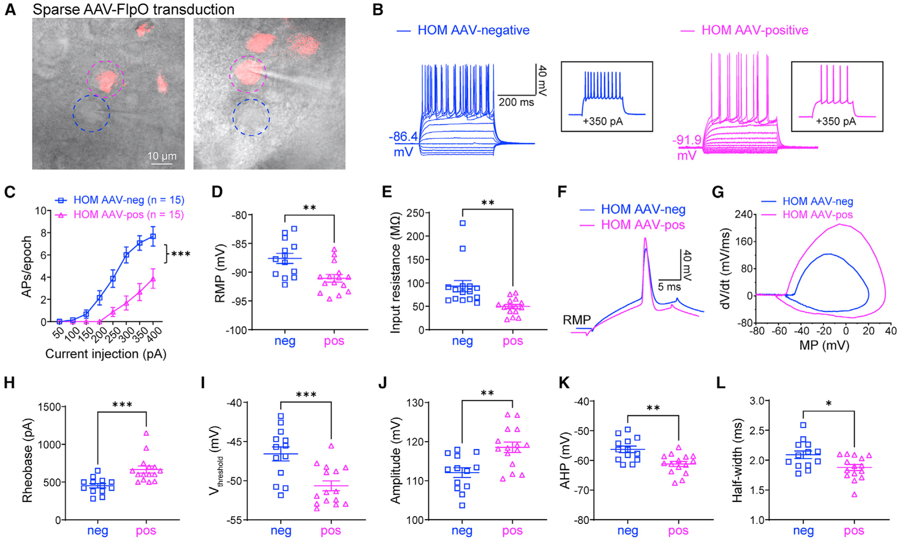
Figure 3. Elevated neuronal excitability is autonomous in adult NaV1.2-deficient mice.

(A) Scn2agt/gt (HOM) mice were injected with a dilute FlpO virus, sparsely transducing a subset of neurons in the striatum. Dashed circles highlight two neighboring AAV-negative (blue circle) and AAV-FlpO-positive (magenta circle) neurons. Scale bar, 10 μm.
(B) Representative current-clamp recordings of AAV-negative (blue) and AAV-FlpO-positive (magenta) MSNs in the CPu of HOM mice were obtained at the RMP. Inset: representative trace in response to +350 pA injection.
(C) The average number of APs generated in response to depolarizing current pulses. Unpaired Mann-Whitney U test for each current pulse.
(D) Individuals and average RMP values. Unpaired t test.
(E) Individuals and average input resistance values at the RMP. Unpaired t test.
(F) Typical spikes were obtained at the RMP.
(G) Associated phase-plane plots.
(H–L) Individual and average spike rheobase, voltage threshold, amplitude, AHP, and half-width values. Unpaired t test. Data are shown as mean ± SEM.
