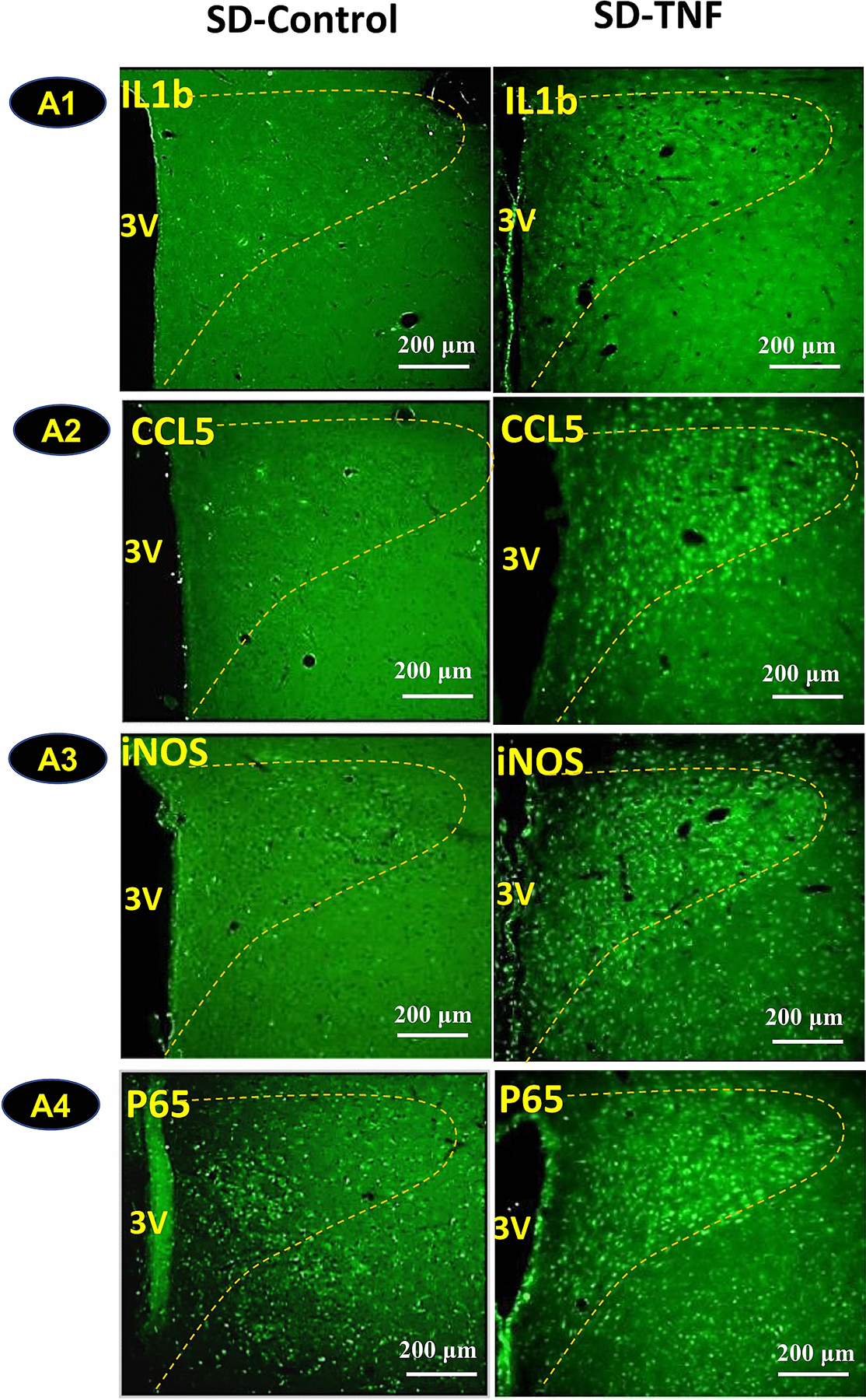
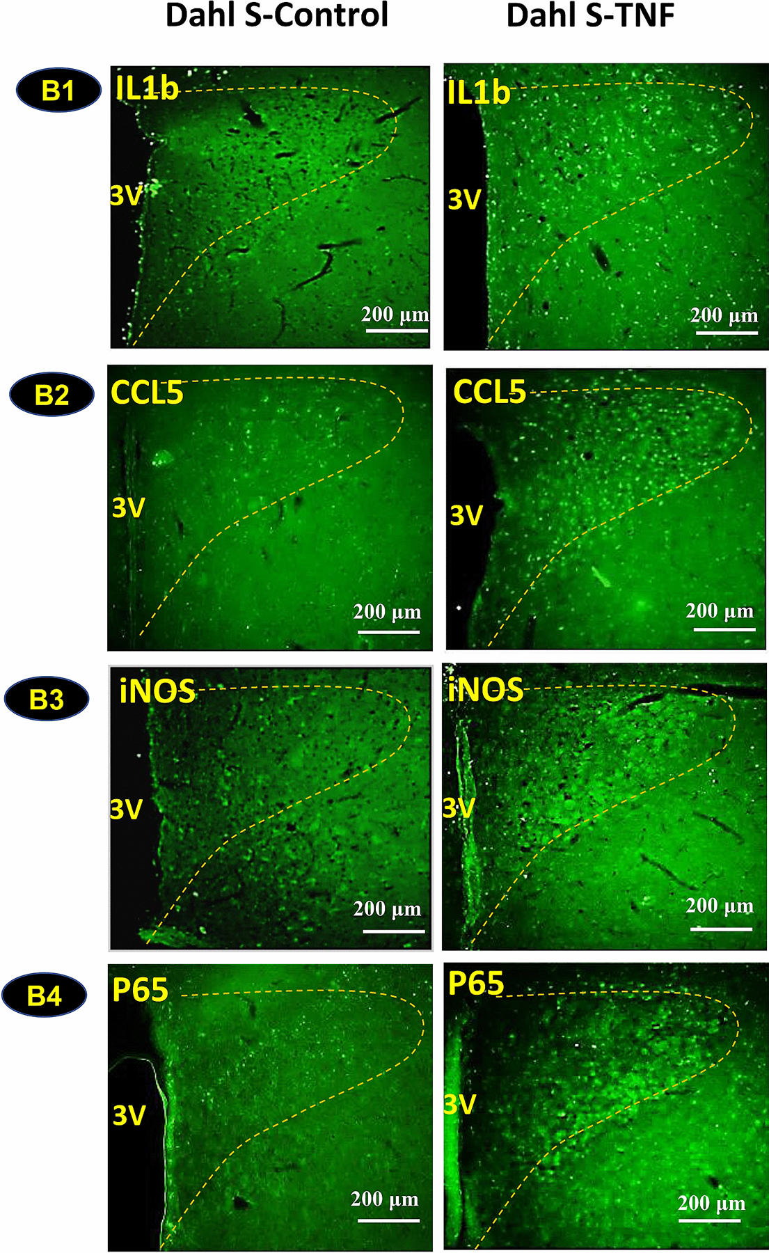
Fig. 4:


Intracerebroventricular (ICV) injection of Tumor Necrosis Factor-α (TNFα) induces increases in inflammatory mediators in the Paraventricular Nucleus (PVN) of Sprague Dawley (SD) rats (A) and Dahl Salt Sensitive (Dahl-S) rats (B). Adult SD and Dahl S rats were divided into two groups of each strain (n=3 per group), and received ICV injection of either TNFα (100 ng/µl, 2.5 µl) or vehicle control (0.9% NaCl, 2.5 µl). Three hours following injection, rats were transcranial perfused with 4% paraformaldehyde (PFA). Brain coronal sections containing the PVN (delineated by the dashed yellow outline) were used to perform immunostaining against primary antibodies anti-Interleukin 1β (IL1β), anti- C-C Motif Chemokine Ligand 5 (CCL5), anti-inducible Nitric Oxide Synthase (iNOS) and anti-P65, a NF-kB subunit. Images were taken with an Olympus BX51 TRF Microscope (Olympus, Japan). Representative images showing immunoactivities of IL1β, CCL5, iNOS, and P65 in SD (A1–A4) and Dahl S rats (B1–B4) in response to ICV injection of TNFα (right panels) or vehicle controls (left panels). (3V: third ventricle, The PVN area included in the image is delineated by the dashed line).
