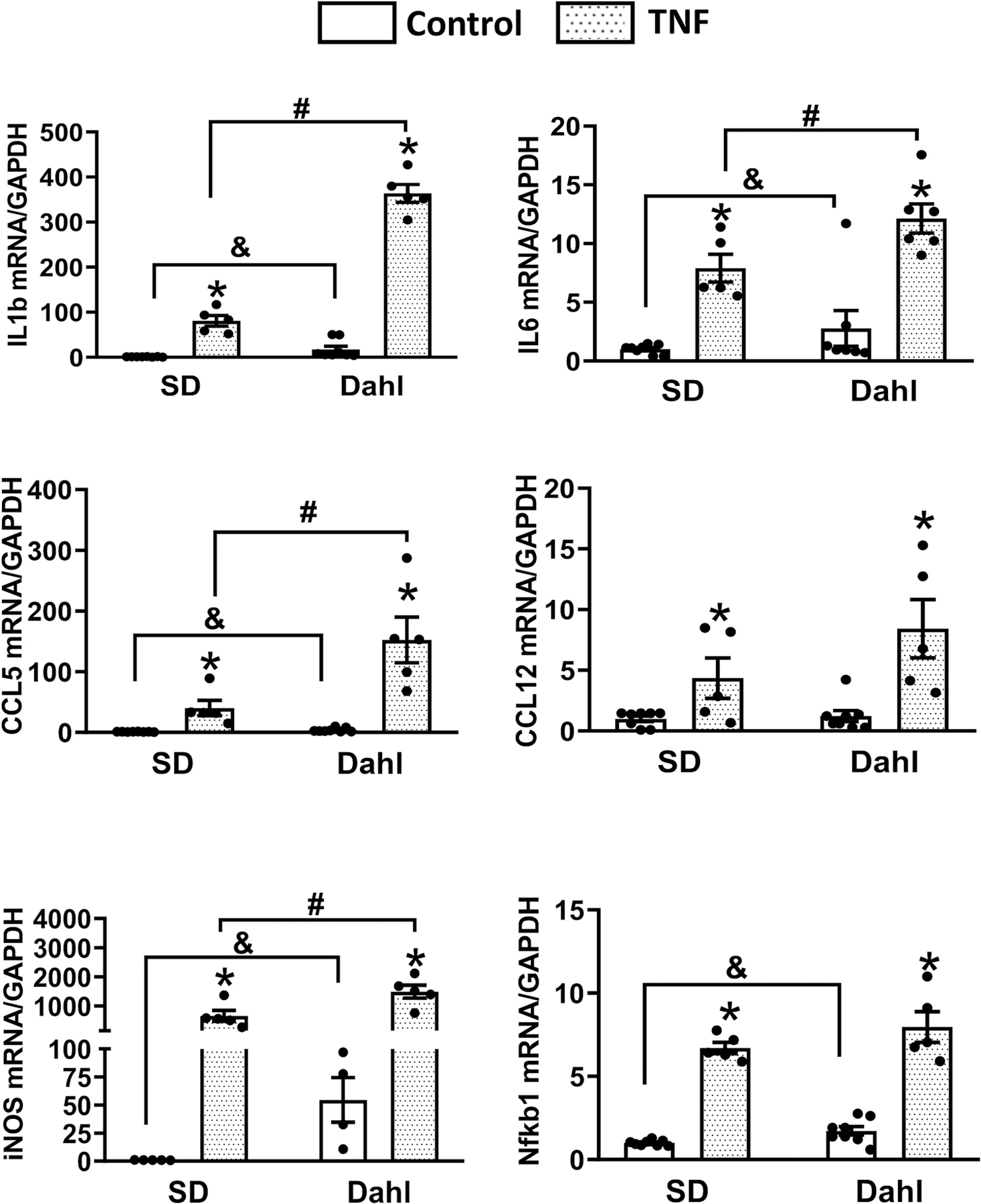
Fig. 5:

Comparison of the inflammatory response in response to Tumor Necrosis Factor-α (TNFα) challenge in the brain neurons from Sprague Dawley (SD) and Dahl-Salt Sensitive (Dahl-S) rats. 10-day-old brain neuronal cultures obtained from the cortex, hippocampus, and hypothalamus of neonatal SD and Dahl-S rats were incubated with either 20 ng/mL TNFα or vehicle control for 6 hours, then cell cultures were collected and subjected to real time Polymerase Chain Reaction (PCR) to measure the mRNA expression of Interleukin 1β (IL1β), Interleukin 6 (IL6), C-C Motif Chemokine Ligand 5 (CCL5), C-C Motif Chemokine Ligand 12 (CCL12), and NF-kappa- p105 (nfkb1). Data were normalized to the housekeeping gene, GAPDH. Each treatment using 2~3 well of neuronal cells, and each experiment was repeated using 3 different batch of cells, the results were combined. (n=6~9/group, *P<0.05 in TNF treatment groups compared to their own control groups; &P<0.05 in Dahl S control group compared to SD control group; #P<0.05 in Dahl-S TNF treatment group compared to the SD TNFα treatment group).
