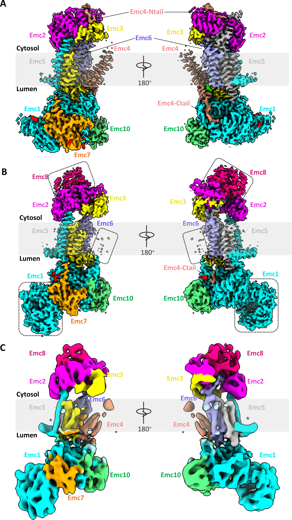
Figure 1. Comparison of the cryo-EM structures of yeast and human EMCs.

(A) Cryo-EM 3D map of the yeast EMC at 3.0-Å overall resolution in front and back views, as viewed from within membrane plane (EMD-21587). (B) Cryo-EM 3D map of the human EMC at 3.4-Å overall resolution (EMD-21929). Dotted rectangles mark unique features in the human EMC: metazoan-specific EMC8 (top), disordered TMD of EMC4 (middle), and NTD1 of EMC1 (bottom). (C) Cryo-EM 3D map of the human EMC at 6.4-Å overall resolution (EMD-11058).
