
Figure 3. Modeling of the yeast Emc4 subunit.
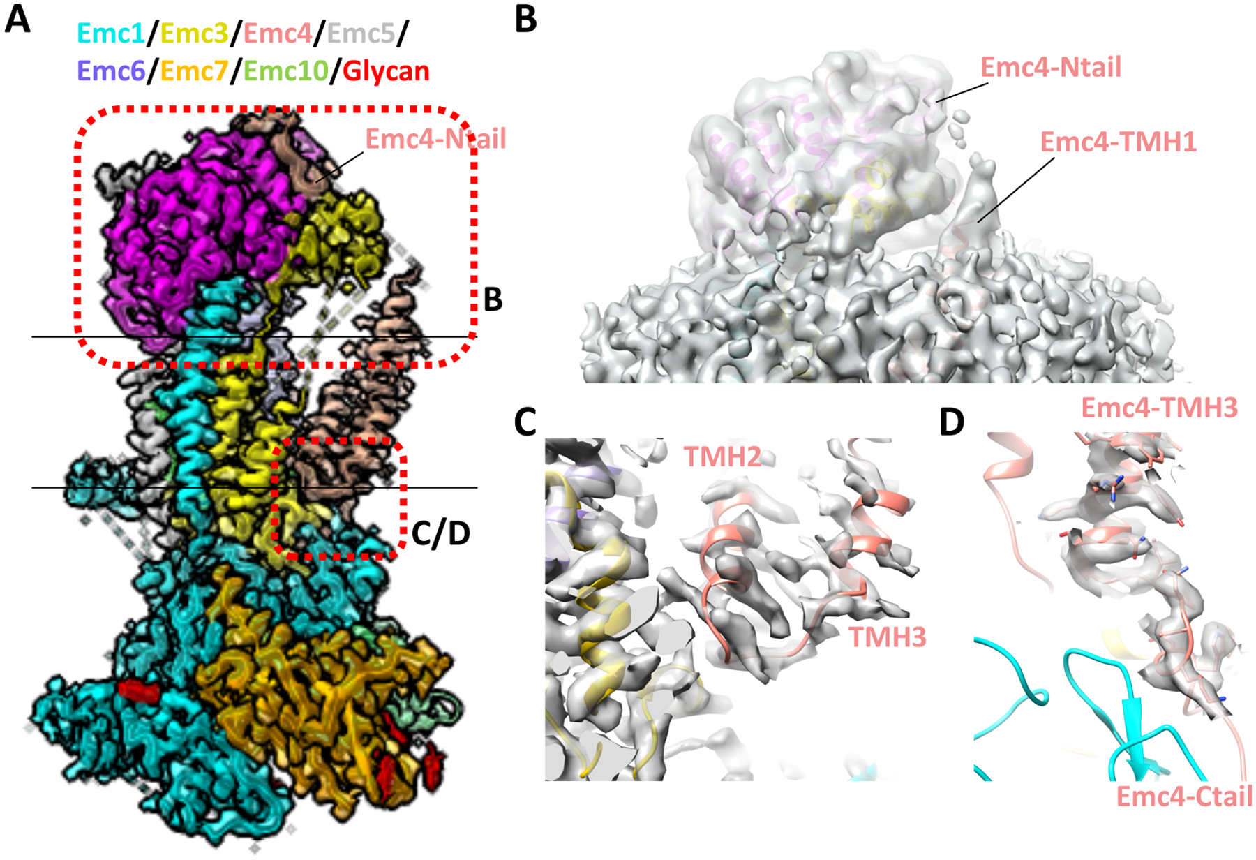
(A) Cryo-EM map of the yeast EMC superposed with the structure shown in cartoon (EMD-21587, PDB ID 6WB9). (B) Linker peptide density between the Emc4 N-tail and TMH1 is shown at a lower threshold. (C) Linker peptide density between Emc4 TMH2 and TMH3. (C) Linker peptide density between Emc4 TMH3 and the C-tail.