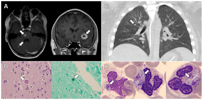
Figure 1.
Clinical and pathological evidence of disseminated toxoplasmosis from the cases presented. (A) Axial and coronal contrast-enhanced magnetic resonance imaging of the brain from case 1 showing rim-enhancing lesions. (B) Representative slides stained with hematoxylin and eosin (HE; left image) and Grocott's methenamine silver stain (right image) showing cysts containing Toxoplasma bradyzoites in the brain tissue from case 1. (C) Axial contrast-enhanced chest computed tomography from case 2 showing bilateral centrilobular nodules and ground glass opacities. (D) Representative slides stained with HE showing neutrophils containing Toxoplasma tachyzoites in peripheral blood from case 2.

