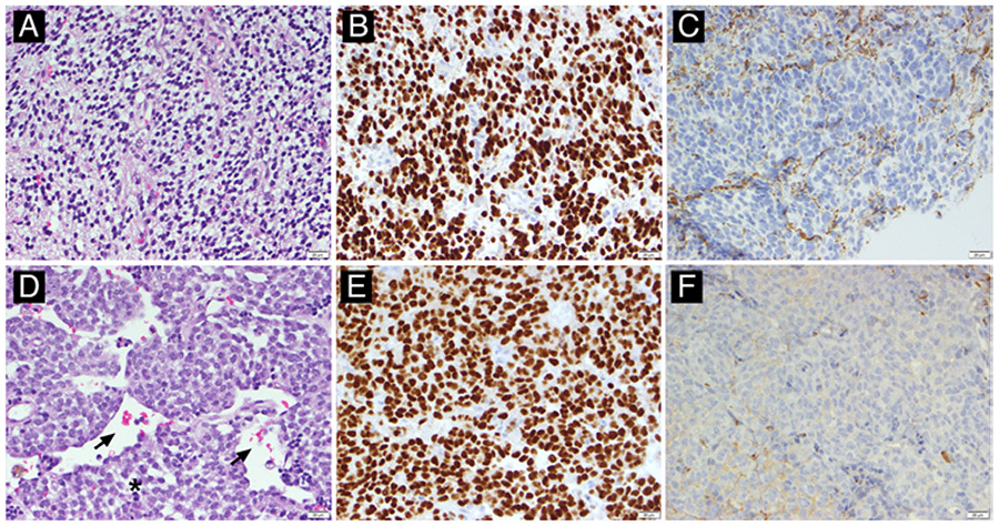
FIG. 1.
Representative histopathology for low- and high-grade canine oligodendrogliomas. A: Low-grade oligodendroglioma (no. 017), H&E stain. The neoplasm is densely cellular, composed of cells with a classic “fried egg” cytoplasmic retraction artifact and bland, monotonous nuclei with minimal atypia. This tumor had only rare mitotic activity, and no geographic necrosis or microvascular proliferation. B and C: Neoplastic cells have diffuse, strong nuclear immunolabeling for oligodendrocyte transcription factor 2 (Olig2; B) and lack immunolabeling for glial fibrillary acidic protein (GFAP; C). D: High-grade oligodendroglioma (no. 011), H&E stain. The neoplasm is densely cellular, with round to ovoid cells aggregated in sheets admixed with minimal stroma, with numerous dilated blood vessels (arrows). In other areas (not pictured), this tumor had striking microvascular proliferation. Cells have variably defined, eosinophilic cytoplasm and plump ovoid or cleaved nuclei with clumped chromatin and 1–2 nucleoli. There are 1–2 mitotic figures (asterisk indicates telophase) per ×40 field, and cells have moderate anisocytosis and anisokaryosis. E and F: Neoplastic cells have diffuse, strong nuclear immunolabeling for Olig2 (E) and lack immunolabeling for GFAP (F). All images were taken at ×400 magnification.

