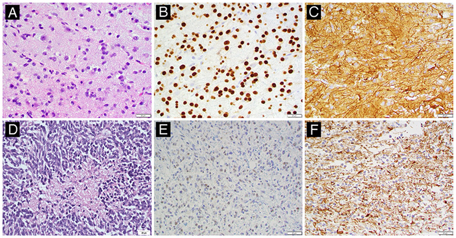
FIG. 2.
Representative histopathology for low- and high-grade canine astrocytomas. A: Low-grade astrocytoma (no. 004). H&E staining reveals a moderately cellular neoplasm consisting of loosely arranged streams and sheets of elongate cells supported by a scant fibrovascular stroma and faintly basophilic mucinous material. Neoplastic cells have mild pleomorphism and indistinct cytoplasmic borders, with a moderate amount of eosinophilic, finely fibrillar cytoplasm. Nuclei are round to elongate and have dense to finely stippled chromatin with 1–2 nucleoli and a low mitotic figure count. B and C: Neoplastic cells exhibit diffuse, strong nuclear immunolabeling for Olig2 (B) and strong cytoplasmic immunolabeling for GFAP (C). D: High-grade astrocytoma (no. 009, necropsy specimen). H&E staining reveals a densely cellular neoplasm consisting of pleomorphic spindloid to polygonal cells that have indistinct cytoplasmic borders. Nuclei are irregularly round to elongate with coarsely stippled chromatin and variably distinct nucleoli. Extensive mitotic figures are present throughout the tumor (28/10 per high-power field) and there are multifocal areas of necrosis and hemorrhage throughout the tumor. E and F: Neoplastic cells exhibit multifocal, weak nuclear immunolabeling for Olig2 (E) and strong cytoplasmic immunolabeling for GFAP (F).

