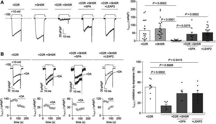
FIGURE 2.
LEAP2 impairs the basal reduction of CaV2.2 currents by GHSR and D2R coexpression and LEAP2 ameliorates the ability of GHSR to impair dopamine-induced inhibition of CaV2.2 currents. (A) Representative traces (left) of CaV2.2 current (ICaV2.2) from HEK293T cells cotransfected with CaV2.2, CaVβ3, CaVα2δ1 and either D2R (+D2R, n = 18), GHSR (+GHSR, n = 21) or GHSR and D2R (+D2R +GHSR, n = 17) pre-incubated or not with 1 µM SPA (+D2R +GHSR +SPA, n = 10) or 0.1 µM LEAP2 (+D2R +GHSR +LEAP2, n = 21) in a 0.1 GPCR:CaV2.2 molar ratio. Bars (right) represent averaged ICaV2.2 levels for each condition. Statistical significance was evaluated by Kruskal-Wallis and Dunn’s post-test. (B) Representative traces and time courses (left) of CaV2.2 current (ICaV2.2) from HEK293T cells cotransfected with CaV2.2, CaVβ3, CaVα2δ1 and D2R (+D2R, n = 9) or GHSR and D2R pre-incubated or not (+D2R +GHSR, n = 7) with 1 µM SPA (+D2R +GHSR +SPA, n = 5) or 0.1 µM LEAP2 (+D2R +GHSR +LEAP2, n = 8) in control condition and after dopamine application (10 µM, +DA); 0.1 GPCR:CaV2.2 molar ratio. Bars (right) represent averaged ICaV2.2 inhibition by 10 µM dopamine application for each condition. Statistical significance was evaluated by Kruskal-Wallis and Dunn’s post-test (versus +D2R). The test-pulse protocol consisted in square pulses applied from −100 to +10 mV for 30 ms every 10 s.

