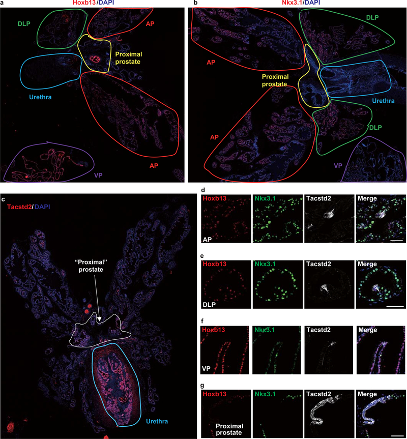
Extended Data Fig. 3 |. Location of Luminal-A, Luminal-B and Luminal-C cells in different prostate lobes.
a,b, Immunofluorescence of Hoxb13 (a) and Nkx3.1 (b) in WT mouse anterior prostate (AP) (red circles), dorsal-lateral prostate (DLP) (green circle), ventral prostate (VP) (purple circle), proximal prostate (yellow circle), and urethra (light blue circle). c, immunofluorescence of Tacstd2 in the whole prostate of T2Y mouse. White circles indicate all the proximal prostate; green circle indicate the urethra. d,e,f,g Co-immunofluorescence of Hoxb13, Nkx3.1 and Tacstd2 in AP (d), DLP (e), VP (f) and proximal prostate (g) of WT prostate. 3 independent mice were used for each experiment. Scale bars, 50 μm(d-g).

