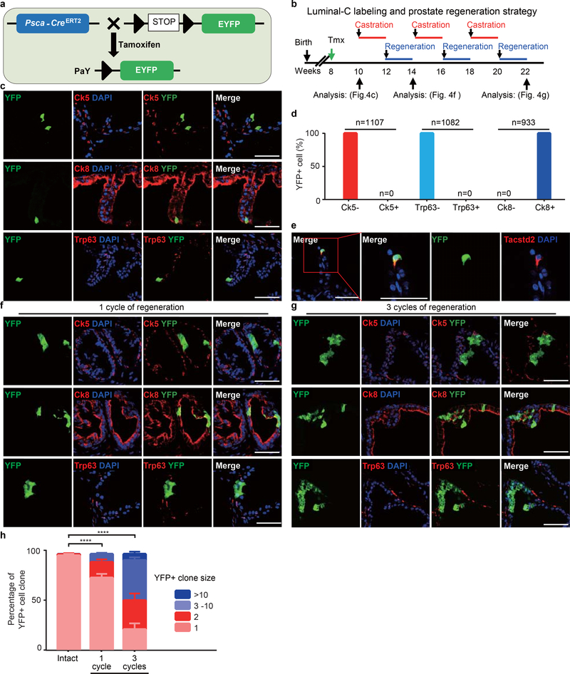
Extended Data Fig. 7 |. Lineage tracing of Psca-expressing Dist-Luminal-C cells.
a, Schematic of targeting strategy to generate the PaY (PscaCreERT2/+; Rosa26EYFP/+) mouse is used to label Psca-expression cells by EYFP expression at 8-week male mice. b, Timeline for Luminal-C cell labeling and prostate regression-regeneration in PaY mouse prostate. c, Co-immunofluorescence of Ck5, Ck8 and Trp63 with endogenous YFP in intact PaY mouse prostates 2-weeks after tamoxifen injection. d, Percentage of YFP+ cell in Ck5− cells, Ck5+ cells, Trp63− cells, Trp63+ cells, Ck8− cells and Ck8+ cells of 10-week PaY mouse prostate. e, Co-immunofluorescence of Tacstd2 with endogenous YFP in intact PaY mouse prostate distal regions 2-weeks after tamoxifen injection. f, Co-immunofluorescence of Ck5, Ck8 and Trp63 with endogenous YFP in regenerated prostate after one cycle of regression-regeneration. g, Co-immunofluorescence of Ck5, Ck8 and Trp63 with endogenous YFP in regenerated prostate after three cycles of regression-regeneration. h, Percentage of YFP-positive cell clone in intact regenerated prostate and regenerated prostate after one or three cycles of regression-regeneration. 8 independent mice were used for each experiment. Scale bars, 50 μm (c, e, f, g). Data show mean ± standard deviation and two-way ANOVA (h). ****P<0.0001 (h)

