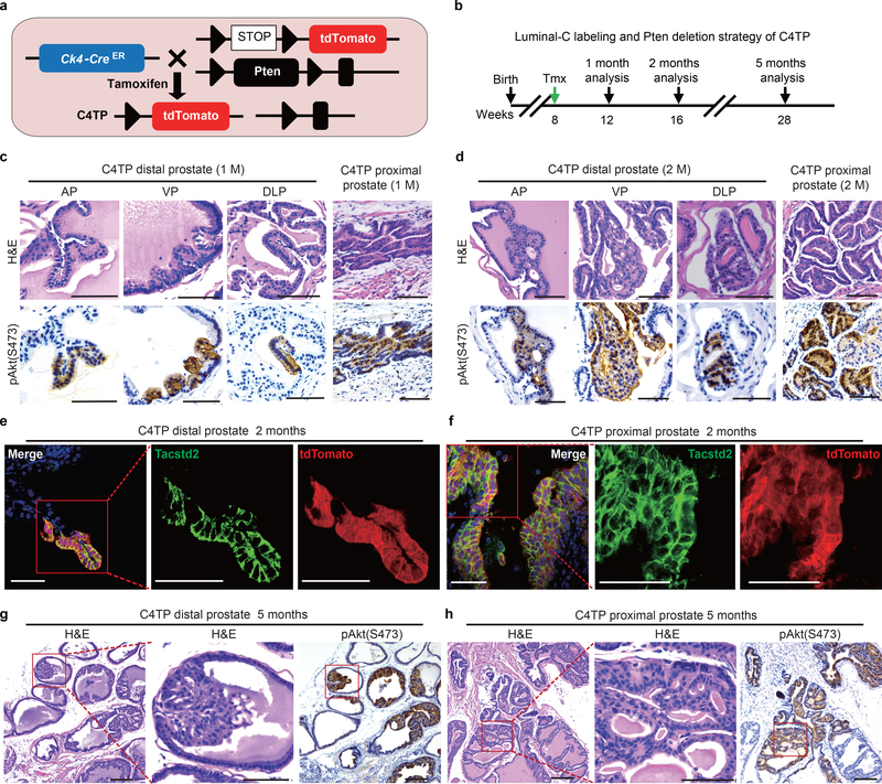
Figure 5 |. Prostate hyperplasia and PIN derived from the Luminal-C cell population.
a, Targeting strategy to generate the C4TP (PscaCreERT2/+;Rosa26EYFP/+;Ptenflox/flox) mouse, label Ck4-expressing cells with tdTomato and perform lineage-specific deletion of the tumor suppressor Pten. b, Schematic of the Luminal-C labeling and Pten deletion strategy in C4TP mice. c,d, Images of H&E staining and pAkt(S473) immunohistochemistry of the C4TP distal prostate at 1 month (c) or 2 months (d) after tamoxifen injection: anterior lobe (left panel), ventral lobe (middle panel), and dorsal-lateral lobe (right panel). e,f Immunofluorescence staining of Tacstd2 and endogenous tdTomato shows that all Luminal-C lineage-marked daughter cells are Tacstd2-positive with Pten deletion. C4TP prostate distal region (left panel) and proximal region (right panel). g,h Images of H&E staining and pAkt(S473) immunohistochemistry of the distal region (g) and proximal region (the square shows the PIN lesion adjacent to the intermediate region) (h) of the C4TP mouse prostate 5 months after tamoxifen injection. Five mice were analyzed for each experiment (c-h). Scale bars, 50 μm (c-h).

