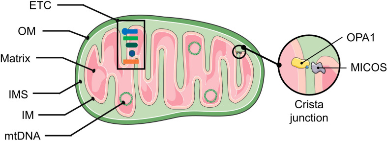
FIGURE 1.
The structure of mitochondria. The mitochondrial double membrane is a tubular reticulum, including an outer membrane (OM) and inner membrane (IM). The OM is porous and freely permits the diffusion of molecules smaller than 5 kD. The IM is a barrier to free ion diffusion and contains several cations and metabolite transporters. The invaginations of the IM that increase the surface area are called cristae. The inner mitochondrial membrane encloses the mitochondrial matrix. An intermembrane space (IMS) exists between the OM and IM, and its composition is close to that of the cytoplasmic matrix. The crista junction represents the architecture by which the cristae are attached to the inner membrane via narrow stems (Panek et al., 2020). The MICOS complex and OPA1 are involved in shaping the cristae. OPA1 act as a tether that maintains “tight” junctions to sequester cytochrome c and oxidative-phosphorylation protein complexes within the crista membrane (Mannella, 2020). Abbreviations: ETC, electron transport chain; mtDNA, mitochondrial DNA; MICOS, mitochondrial contact site and cristae organization system.

