FIGURE 2.

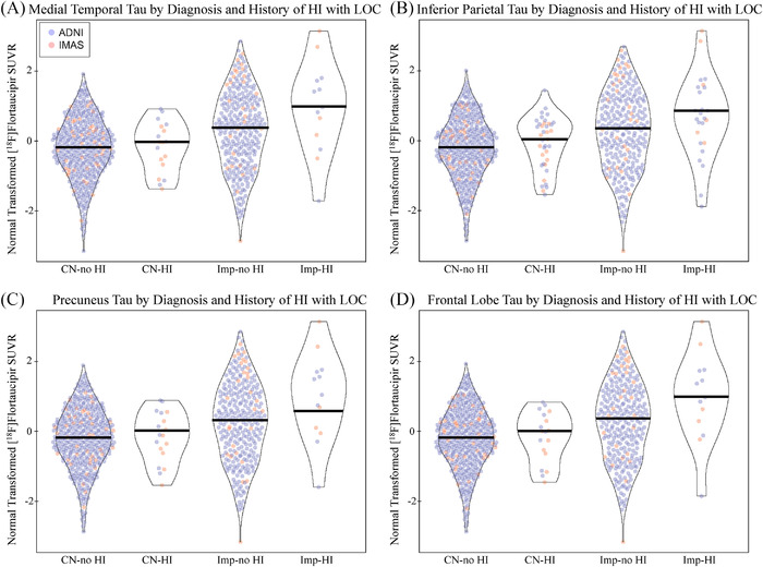
Tau deposition by diagnosis and history of head injury with loss of consciousness. Individuals with a history of head injury (HI) with a loss of consciousness (LOC) show significantly greater normal transformed [18F]flortaucipir SUVR in the (A) medial temporal lobe (DX: P < .001, d = 0.296; HI with LOC: P = .049, d = 0.144; DX by HI with LOC: P = .104, d = 0.215), (B) inferior parietal lobe (DX: P < .001, d = 0.295; HI with LOC: P = .025, d = 0.164; DX by HI with LOC: P = .096, d = 0.220), (C) precuneus (DX: P < .001, d = 0.294; HI with LOC: P = .036, d = 0.154; DX by HI with LOC: P = .083, d = 0.230), and (D) frontal lobe (DX: P < .001, d = 0.296; HI with LOC: P = .034, d = 0.149; DX by HI with LOC: P = .086, d = 0.227). This effect appears to be due to increased tau in impaired participants with HI with LOC. Age, sex, mean global amyloid, and cohort were included as covariates. Note: Participants include 432 CN without history of HI with LOC, 18 CN with history of HI with LOC, 289 impaired without history of HI with LOC, 13 impaired with history of head injury with LOC. Abbreviations: ADNI, Alzheimer's Disease Neuroimaging Initiative; CN, cognitively normal; d, Cohen's d; DX, diagnosis; HI, head injury; IMAS, Indiana Memory and Aging Study; LOC, loss of consciousness; SUVR, standardized uptake value ratio
