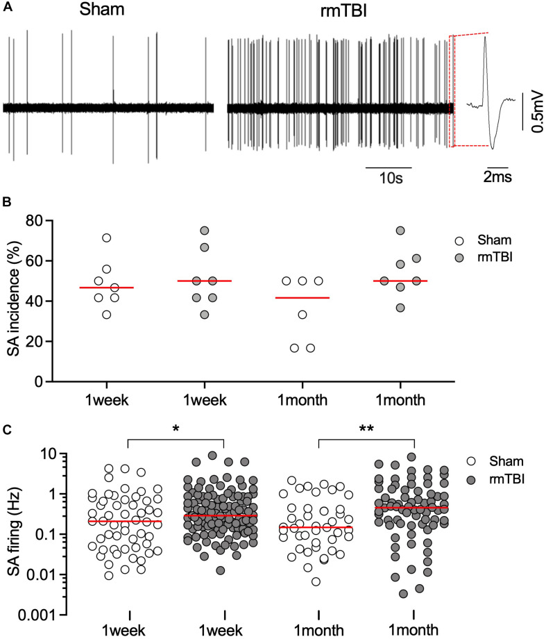
FIGURE 8.
rmTBI on excitatory neuronal activity in mPFC. (A) Sample recordings from rat 1 months after sham injury (left) and rmTBI (right). (B) There is no difference in incidence of spontaneous firing between sham injury and rmTBI groups. Each dot represents recording from one rat. (C) Firing rates of spontaneous firing of mPFC pyramidal neurons are increased after rmTBI. Each dot represents data from one neuron recorded (n = 6–7 rats per group showing in B). Red bars represent means. *P < 0.05, **P < 0.01 by the planned comparison with Bonferroni correction.

