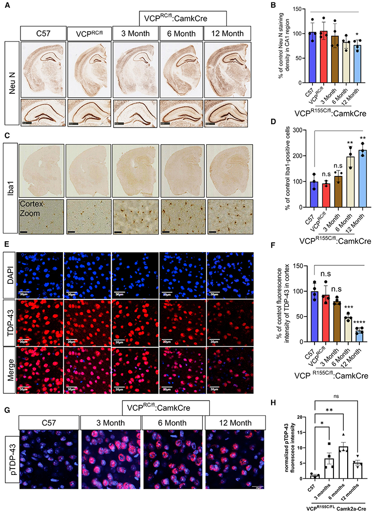
Figure 5. Re-expression of a VCP-R155C mutant allele in VCP cKO mouse neurons recapitulates FTLD-TDP pathology.

(A) NeuN staining of coronal sections through the cortex and hippocampus of 9-month-old control (C57), 9-month-old VCP-R155C heterozygous (VCPRC/FL), or VCP cRC (VCPRC/FL:CamkCre) mice at 3, 6, and 12 months of age. Scale bars, 500 μm in bottom panel.
(B) Quantitation of NeuN. Data represent mean ± SD (error bars) (n = 3 slices from four mice per group). *p < 0.05 with paired t test against C57 control.
(C) Iba1 staining of coronal sections through the cortex and hippocampus of 9-month-old control (C57), 9-month old VCP-R155C heterozygous (VCPRC/FL), or VCP cRC (VCPRC/FL:CamkCre) mice at 3, 6, and 12 months of age. Scale bars, 30 μm in bottom panel.
(D) Quantitation of Iba1 staining. Data represent mean ± SD (error bars) (data points represent mice). A one-way ANOVA was used for statistical testing, treating three slices from three mice as an independent sample; F(4,10) = 14.83, p = 0.0003. Post hoc comparisons using Tukey’s test, **p < 0.01.
(E) TDP-43 (red) immunofluorescence and DAPI nuclear (blue) fluorescence of the cortex from 9-month-old control (C57), 9-month-old VCP-R155C heterozygous VCPRC/FL), or VCP cRC (VCPRC/FL:CamkCre) mice at 3, 6, and 12 months of age. Scale bars, 30 μm.
(F) Quantitation of TDP-43 fluorescence intensity. Data represent mean ± SD (error bars) (n = 3 slices from four mice per group). Data represent mean ± SD (error bars) (data points represent mice). A one-way ANOVA was used for statistical testing, treating three slices from four mice as an independent sample; F(4,15) = 32, p < 0.0001. Post hoc comparisons using a Bonferroni test, ***p < 0.0001.
(G) Phospho-TDP-43 (red) immunofluorescence and DAPI nuclear (blue) fluorescence of the cortex from 9-month-old control (C57) or VCP cRC (VCPRC/FL:CamkCre) mice at 3, 6, and 12 months of age. Scale bar, 20 μm.
(H) Quantitation of pTDP-43 fluorescent intensity. Data represent mean ± SD (error bars) (n = 3 slices from four mice per group). F(3,11) = 8.469, p = 0.0034 followed by Tukey’s test was used for comparisons.
