FIGURE 1.
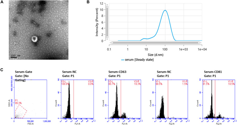
Ultrastructure and characterization of serum-derived exosomes. (A) TEM image shows that the exosomes have a cup-shaped structure. Bar, 100 nm. (B) Of the particles, 89.026% were 20–200 nm in size, as detected by nanoparticle tracking analysis (NTA). (C) Flow cytometry analysis of the exosome markers CD63 and CD81, with 19.3 and 18.5% positive rates, respectively.

