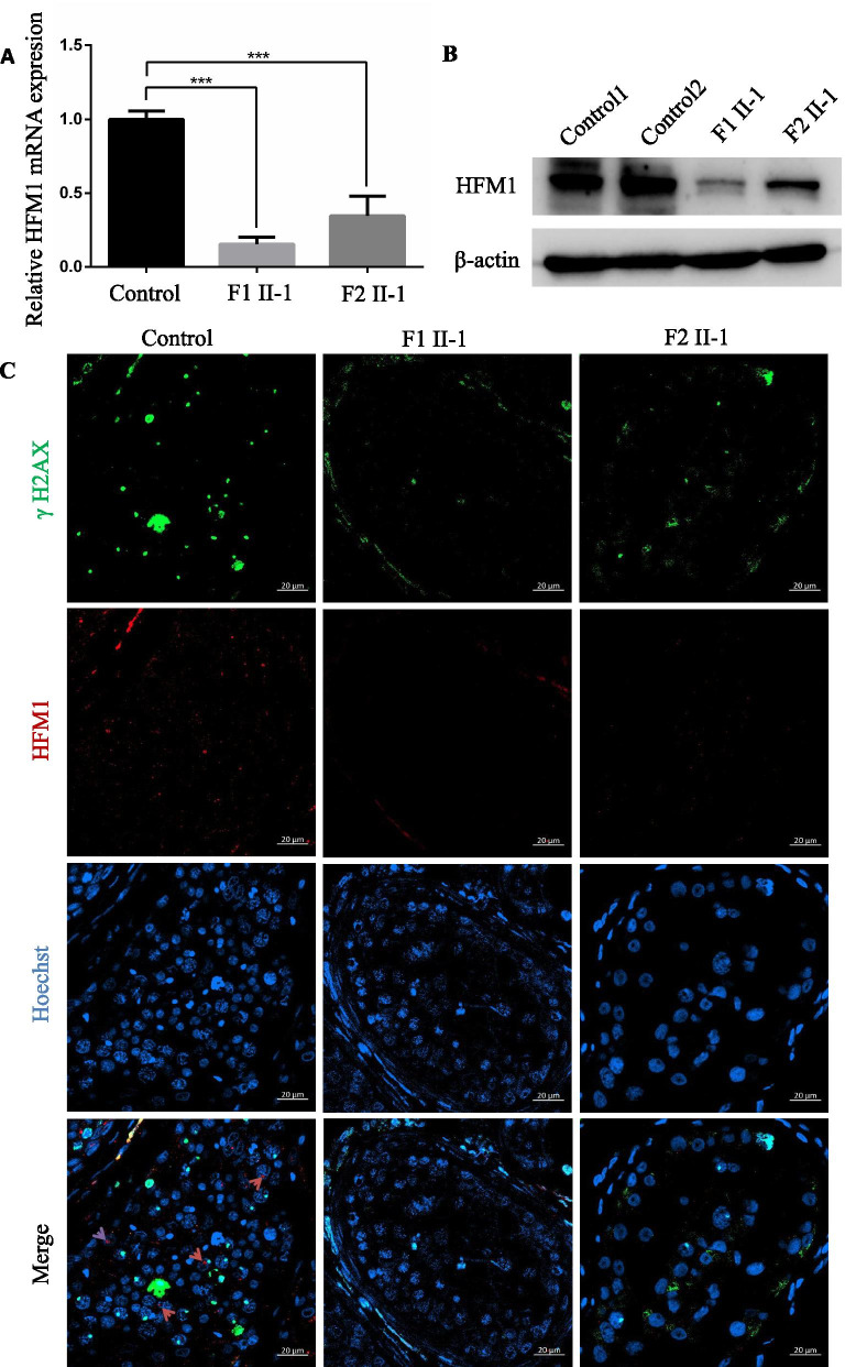
Fig. 5.
Expression and location analysis of HFM1 protein in the testis from the control patient and men harboring bi-allelic HFM1 variants. A qPCR analysis indicated that the abundance of HFM1 mRNA decreased significantly in the testis of men harboring homozygous HFM1 variants when compared to that of a normal control male. Data represents the means ± SEM. (standard error of measurement) of three independent experiments. Two-tailed Student’s paired or unpaired t tests were used as appropriate (*** P < 0.001). B Western blotting assay revealed that HFM1 protein levels reduced significantly in the testis from men harboring HFM1 mutations. β-actin was used as a loading control. C HFM1 localization in the testis from a control individual and men harboring bi-allelic HFM1 variants. HFM1 immunostaining (red) was primarily concentrated in cytoplasm of spermatogonia and spermatocytes in seminiferous tubules in normal control. The immunostaining was decreased in the testicular tissues of F1 II-1 and F2 II-1. Hoechst (blue) was stained as a nuclear marker. The γH2AX (green) was stained as a marker of spermatocyte. Scale bars: 20 μm

