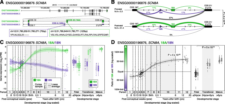
Fig. 5.
Developmental trajectories of CDS 20 (18A/18N) in human cortex in SCN8A. A Location, genomic coordinates (GRCh38/hg38), and amino acid sequence of the 18A and 18N exons in SCN8A. B Sashimi plot of intron splicing in prenatal (top, N = 112 samples) and postnatal (bottom, N = 60 samples) dorsolateral prefrontal cortex. Linewidth reflects the proportion of split reads observed for each intron compared to all split reads between the exon before and after, this value is also shown as a percentage. Introns related to 18A exon inclusion are shown in green, those related to 18N exon inclusion are shown in purple, and others are in grey. C Expression of the 18A (green) and 18N (purple) for 176 BrainVar human dorsolateral prefrontal cortex samples across development (points). On the left, the colored line shows the Loess smoothed average with the shaded area showing the 95% confidence interval. On the right, boxplots show the median and interquartile range for the same data, binned into fetal, transitional, and mature developmental stages. D The 18A:18N ratio is shown for each sample from panel C across development (left) and binned into three groups (right). CPM: Counts per million; Statistical analyses: B Dirichlet-multinomial generalized linear model, as implemented in Leafcutter [41]. D Left panel, linear regression of log2(18A:18N ratio) and log2(post-conceptual days). Right panel, two-tailed Wilcoxon test of log2(18A:18N ratio) values between fetal and mature groups

