Figure 9.

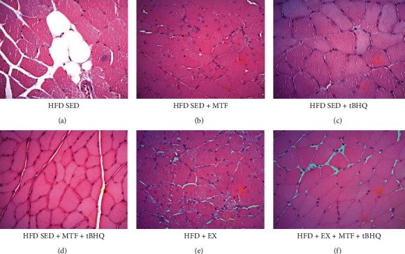
Gastrocnemius histology of HFD-treated groups. Crosssectional views of gastrocnemius stained with H&E are shown to differentiate structure and arrangement of muscle fibers obtained for the HFD rats after the different interventions at 15 months of age. Micrographs are 40x magnification. n = 3.
