Figure 2.
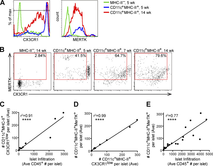
MERTK-expressing cells increase with progression of islet infiltration. Islets from 4–18-wk-old NOD or NOD.CX3CR1-GFP female mice were isolated, digested and dissociated, stained, and analyzed by flow cytometry. (A) Example histograms of CX3CR1 gating in female NOD.CX3CR1-GFP islets. (B) Example plots of MERTK+CX3CR1Low gating in female NOD islets, with percentage of positive in the gate shown. (C–E) Each dot represents one mouse, as the number of cells in the islets normalized to the number of islets per mouse (Ave). (C) Quantification of CD11c+MHC-II+CX3CR1ow (infiltrating APCs) versus the leukocyte infiltration in islets. (D) Quantification of CD11c+MHC-II+MERTK+ cells versus CX3CR1low CD11c+MHC-II+ infiltrating APCs in islets. (E) MERTK+CD11c+MHC-II+ versus leukocyte infiltration in islets. n = 11 mice from two independent experiments (A–D); n = 24 mice from six independent experiments (E). Statistics calculated by Pearson correlation; ****, P < 0.0001 (C–E).

