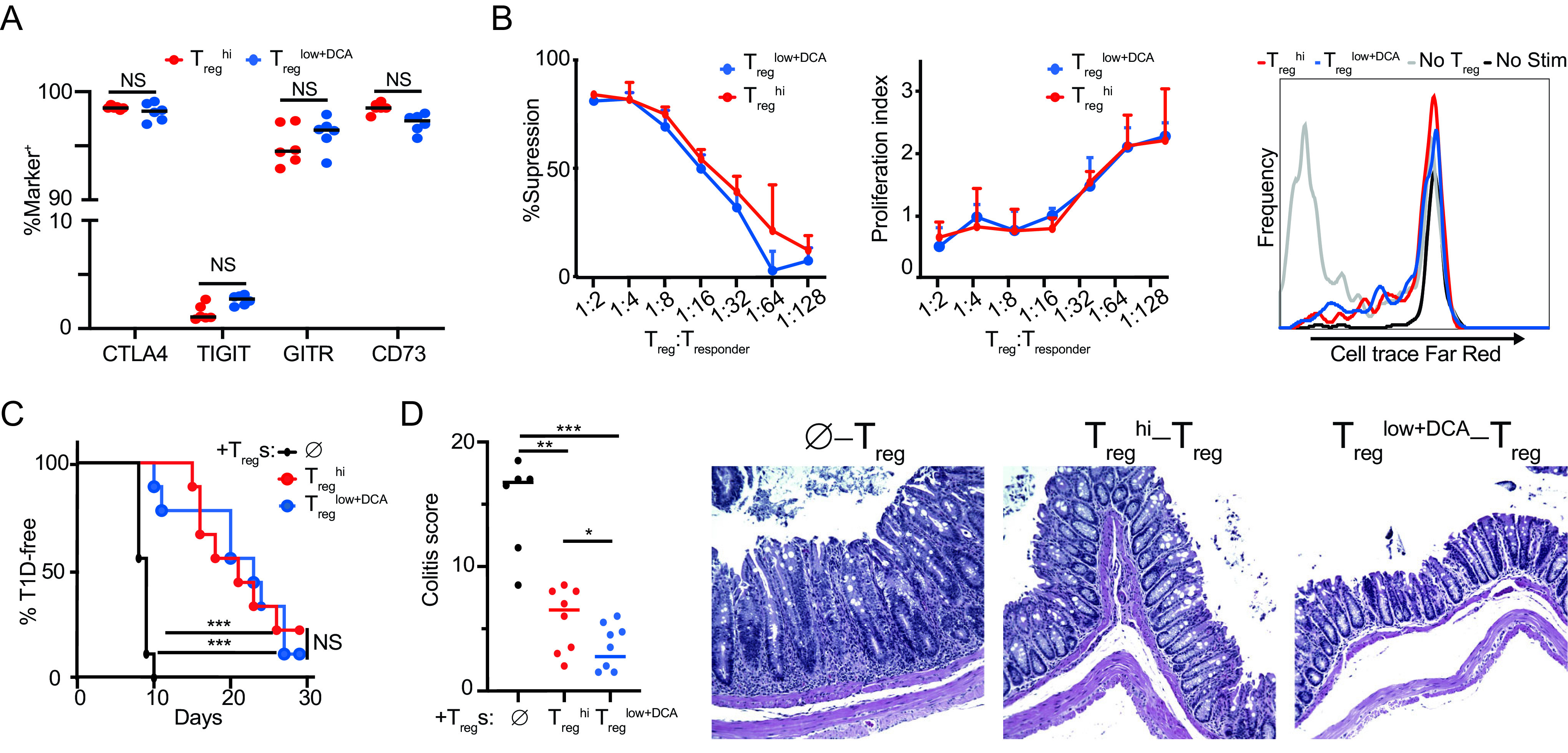
FIG 3.

DCA enhances differentiation of functional Tregs. Phenotype and suppressive function of DCA-driven Tregs (Treglow+DCA) compared to Treghi-driven Tregs. (A) Expression of canonical Treg function-associated proteins. (B) Standard in vitro suppression assay (Tregs at 1:16) showing percentages of suppression (left), proliferation index (middle), and representative histograms (right) (n = 3 technical replicates, representative of 3 experiments; no difference at all points by Student’s t test). (C) NOD.BDC2.5 model of type 1 diabetes (n = 9 mice per condition across 2 experiments). (D) B10.RAG2−/− model of colitis (n = 7 or 8 mice per condition across 2 experiments). , no-Treg controls. Mann-Whitney (A, D) and Mantel-Cox (C) tests were used, *, P < 0.05; **, P < 0.01; ***, P < 0.001; NS, not significant.
