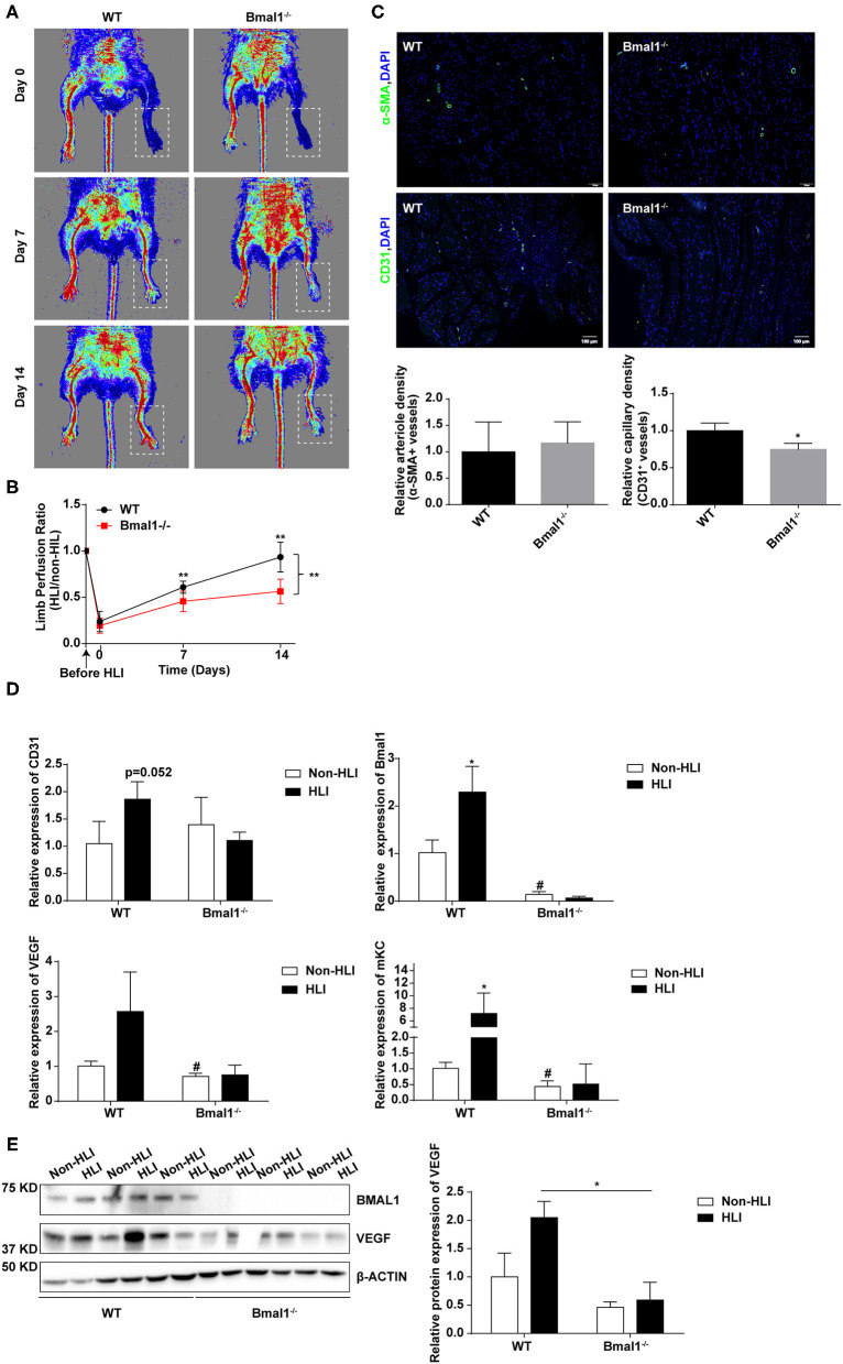
Figure 5.
Bmal1 promotes angiogenesis after peripheral ischemic injury. (A) Blood flow obtained via laser Doppler perfusion imaging on days 0, 7, and 14 after HLI in Bmal1−/− mice and WT mice. (B) The perfusion of the hindlimbs of mice at each time point was calculated as the ratio of measurements of the injured (HLI) and uninjured (non-HLI) limbs. n = 4 for Bmal1−/− and WT mice. Data are presented as mean ± SEM (unpaired t-test and two-way ANOVA with post-hoc Sidak test). **P < 0.01 Bmal1−/− vs. WT. (C) At 14 days after HLI, the gastrocnemius muscle was harvested from the HLI limb of Bmal1−/− mice and WT mice and stained for α-smooth muscle actin (α-SMA) and CD31 expression. Data are presented as mean ± SEM (n = 4, unpaired t-test). *P < 0.05 Bmal1−/− vs. WT. (D) mRNA levels of the CD31, Bmal1, VEGF, and murine functional IL-8 homolog keratinocyte-derived chemokine (mKC) were evaluated in HLI and non-HLI limbs via real-time PCR. Data are presented as mean ± SEM (n = 4, unpaired t-test). *p < 0.05 wild-type (WT) HLI vs. WT non-HLI; #p < 0.05 Bmal1−/− non- HLI vs. WT non-HLI. (E) Protein expression level of BAML1 and VEGF in HLI and non-HLI limbs measured by western blot. Data are presented as mean ± SEM (n = 3, unpaired t-test). *p < 0.05 Bmal1−/− HLI vs. WT HLI.

