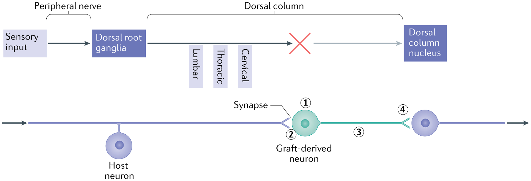
Fig. 2 |. Forming a relay using neural progenitor cells.

The schematic illustrates the elements required to use neural progenitor cells (NPCs) to form a relay following spinal cord injury, using the example of the sensory system in the presence of a cervical lesion of the dorsal column and transplants of neuronal-restricted precursors (NRPs) and/or glial-restricted precursors (GRPs). The upper part illustrates the elements of the ascending sensory pathway, disrupted by a cervical injury that interrupts the connectivity of sensory axons (dorsal column) with the dorsal column nucleus (DCN) in the brainstem. The lower part shows the steps required to restore the connectively using NPC transplants that form a relay. First, the transplant must survive and generate neurons with the appropriate phenotype (excitatory, for example) (1). The use of a mixture of NRPs and GRPs has been found to be effective, as the GRP-derived astrocytes generate a permissive environment for survival and differentiation of neurons16. Second, the host axons must grow into the graft and form synaptic connections (2). It appears that the presence of astrocytes in the graft attracts the sensory neurons40, but other strategies include the induction of the growth potential of host neurons through the repression of genes such as PTEN and SOCS3 (REF.238). Finally, the axons of transplanted neurons must undergo directional extension to the target (along a neurotrophic gradient to the DCN101) (3) and form synaptic connections (4). To verify the formation of a functional relay, analysis needs to be performed at different levels. Structural analysis includes the tracing of axon growth from the host and the transplant and obtaining evidence of synaptic structure by electron microscopy. Physiological analysis may involve the stimulation of axons followed by assessment of the expression of FOS in downstream neurons as well as measures of signal transmission through the transplant. Functional analysis will include behaviour tests indicative of restored connectivity of the specific tracts.
