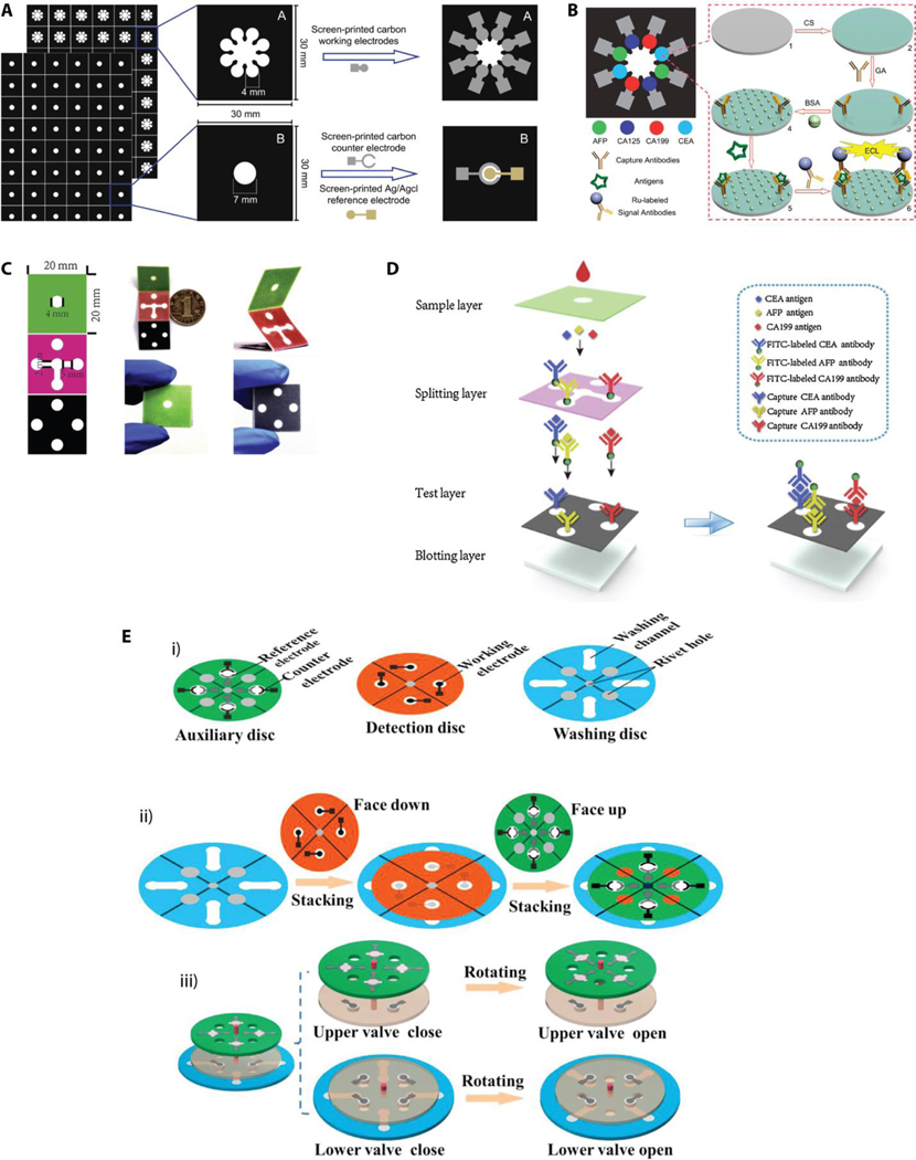
Figure 14.
Schematics of typical multiplex 3D μPADs configurations. a) Wax printed two paper sheets (paper A and paper B) were chemically modified to develop multiple working electrodes on paper A and counter and reference electrodes on paper B. b) Multiple detection zones were dedicated for electrochemical-based detection of four protein biomarkers. Reproduced with permission.[99] Copyright 2012, Elsevier. c) Optical images of the 3D μPAD that exhibits origami style device assembly. d) Description of assay reagents that are loaded on each layer and the flow direction of the sample once applied to the sample layer. Reproduced with permission.[102] Copyright 2020, Elsevier. e) The 3D μPAD including a rotational disks format. i) Three layers of rotating disks with electrodes integrated in the auxiliary and detection disks. ii) The assembly of the rotational 3D device. iii) The control mechanism of the “on/off’ states of upper and lower valves. Reproduced with permission.[103] Copyright 2018, Elsevier.

