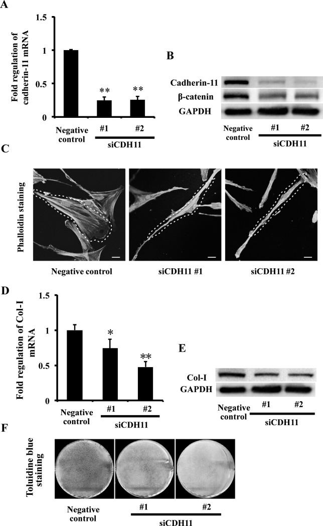
Figure 4.
Knockdown of CDH11 altered PDLC morphology and repressed Col-I expression. (A, B) The mRNA and protein expressions of cadherin-11 and protein expression of β-catenin in PDLCs were suppressed in the siCDH11 No.1 and siCDH11 No. 2 groups, examined by real-time PCR (A) and Western blot (B). ** P = .01 vs negative control group. (C) Phalloidin staining showed morphology of PDLCs in negative control, siCDH11 No.1, and siCDH11 No. 2 groups. Scale bars: 20 μm. (D, E) The mRNA and protein expressions of Col-I in PDLCs were repressed in the siCDH11 No.1 and siCDH11 No. 2 groups, examined by real-time PCR (D) and Western blot (E). * P = .05, ** P = .01 vs negative control group. (F) Toluidine blue staining showed that collagen matrix of PDLCs was repressed in the siCDH11 No.1 and siCDH11 No. 2 groups compared with the negative control group.

