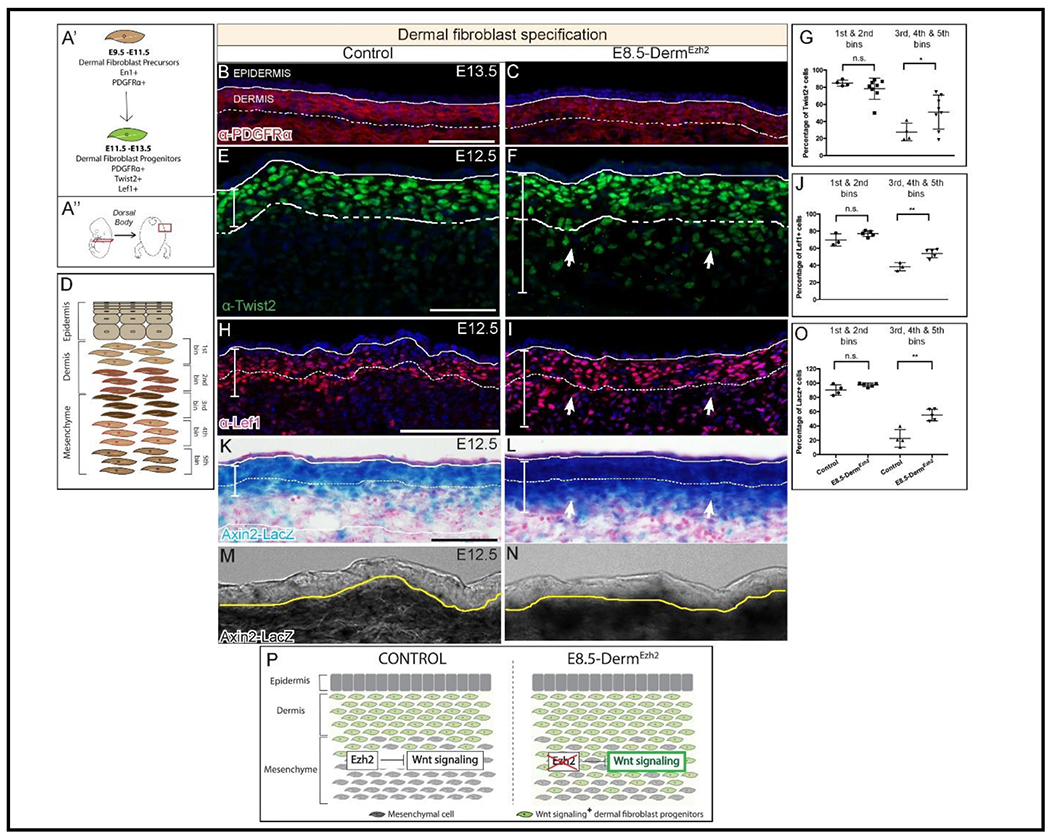
Figure 2: Ezh2 restricts the specification of Wnt/ β-Catenin Signaling mediated DF progenitors to the dermis:

Schematic illustration of the temporal window (A’) and region of interest (A”). Indirect immunofluorescence with DAPI counterstain showing the expression of mesenchymal marker, PDGFRα (B, C), and dermal fibroblast progenitor markers TWIST2 (E, F), and LEF1 (H, I). Axin2-LacZ (β-galactosidase) staining (K, L) at E12.5 along with a digital zoom of a transmitted light channel using a confocal microscope (M, N). Schematic illustration of binning of dermis and dorsal mesenchyme employed to quantify cells (D). Quantification of TWIST2+ (G), LEF1+ (J), and β-galactosidase+ (O) cells in both controls and mutants. A summary schematic depicting the extent of Wnt/β-Catenin signaling domain in control and expansion in the E8.5-DermEzh2 mice (P). Solid line demarcates the epidermis from dermis and dashed line demarcates the lower limit of the dermis. Scale bar=100 microns in B, C, H, I, K, L and 50 microns in E and F.
