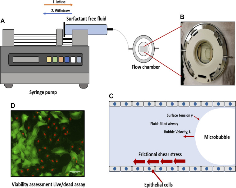
Figure 1.
Experimental setup. A: a single bubble is propagated over cell monolayer by filling the chamber and then retracting fluid over the cells. B: a propagating bubble is seen. C: we exposed epithelial cells to frictional shear stress relevant to airway reopening in our setup. D: fluorescence live/dead stain method was used to assess viability. Here, green is calcein-stained live cells, and red is ethidium-stained dead cells.

