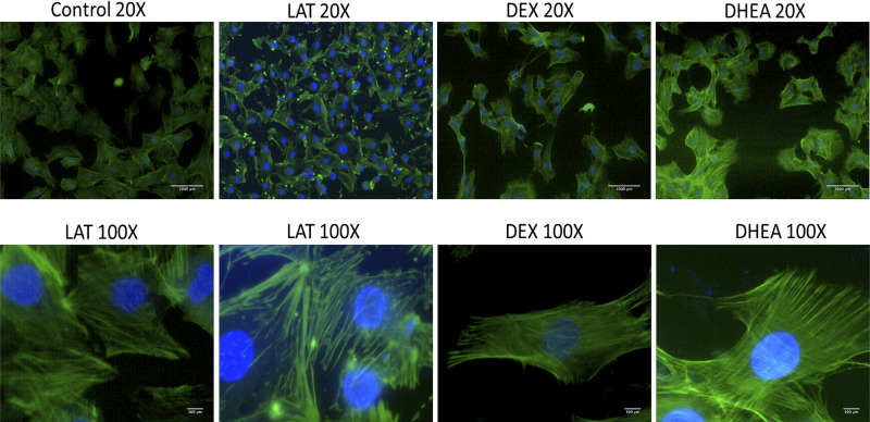
Figure 4.
Immunostaining of actin cytoskeleton for nontreated and treated cells. In the slides, green is phalloidin-stained actin fibers, and blue is DAPI-stained cell nuclei. Compared with controls, all groups did not have any clear difference in the intensity of green, fluorescence signal, except LAL, where actin fibers are depolymerized. For both DEX- and DMSO-treated cells, actin fibers look longitudinally elongated. Scale bar in ×20 and ×100 pictures is 1 mm and 100 µm, respectively. DEX, dexamethasone.

